

 vi
vi 16-Jun-2026
 24,700
 Overview of Vision Impairment
Vision impairment is a condition characterized by a reduced ability to see to some degree, causing difficulties in daily activities that cannot be fully corrected with conventional vision aids such as eyeglasses or standard contact lenses.
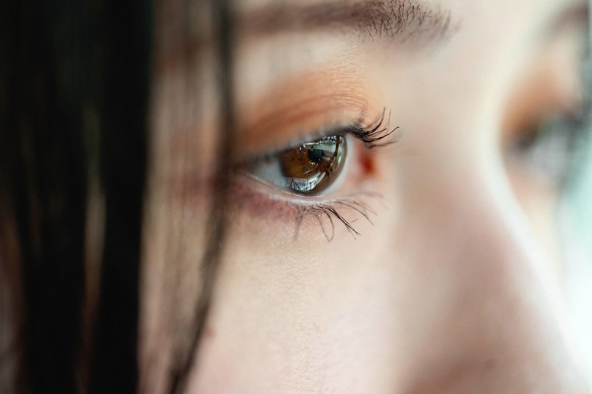
Causes of vision impairment and treatment solutions
Pathophysiology and Mechanisms of Vision Loss
The visual system functions through the following mechanism: Light passes through the cornea and crystalline lens and is precisely focused onto the retina (the light-sensitive neural tissue located at the back of the eye). There, photoreceptor cells convert light signals into nerve impulses, which are transmitted through the optic nerve to the brain for image processing.
Vision impairment occurs when one or more disruptions affect this visual pathway:
- Refractive errors: Light fails to focus properly on the retina due to abnormal eyeball length or irregular corneal curvature.
- Opacity of transparent ocular media: The crystalline lens or vitreous body becomes cloudy, preventing light from reaching the retina.
- Retinal and optic nerve diseases: Damage to signal-receiving or signal-transmitting cells, retinal detachment, or retinal degeneration can distort or completely interrupt visual information reaching the brain.
Classification of Vision Impairment Severity
According to the standard Snellen visual acuity chart, normal vision is defined as 20/20. For cases involving irreversible visual dysfunction, medicine classifies vision impairment into the following categories:
- Low vision: Patients retain some usable vision but experience difficulty performing tasks such as reading or driving. Low vision does not include total blindness.
- Partial vision loss: Visual acuity ranges between 20/40 and 20/200 despite optimal corrective lenses.
- Legal blindness: Visual acuity is no better than 20/200 in the better-seeing eye after correction, or the visual field is restricted to less than 20 degrees. This is a legal definition and does not necessarily mean total blindness (complete inability to perceive light).
Common Causes of Vision Impairment
The causes of vision impairment are highly diverse, ranging from school-age refractive errors to serious retinal diseases commonly seen in older adults.

Common causes of vision impairment include refractive errors
1. Refractive Errors and Presbyopia
These are the most common causes of vision impairment worldwide and can often be effectively managed if detected early:
Myopia (Nearsightedness)
Myopia occurs when images are focused in front of the retina, typically due to an elongated eyeball or excessive corneal refractive power. As a result, distant objects appear blurry. This condition affects males and females equally, usually develops during childhood, and tends to stabilize during the first two decades of life.
Hyperopia (Farsightedness)
In contrast to myopia, images are focused behind the retina, causing nearby objects to appear blurry. Children may naturally have mild physiological hyperopia, which often improves as the eyeball lengthens during growth.
Astigmatism
Astigmatism may occur alone or in combination with myopia or hyperopia when the cornea or crystalline lens has an irregular shape (similar to an oval rugby ball rather than a perfectly round soccer ball). Consequently, the eye cannot focus light onto a single point, resulting in blurred and distorted vision at all distances.
Presbyopia
Presbyopia is the age-related decline in the eye’s natural focusing ability, typically beginning around age 40. People with presbyopia often require reading glasses or must hold reading materials farther away to see clearly. Although presbyopia and hyperopia both affect near vision, presbyopia is caused by reduced elasticity of the crystalline lens and ciliary muscles rather than the length of the eyeball.
2. Disorders of the Ocular Surface and Anterior Segment
Cataracts
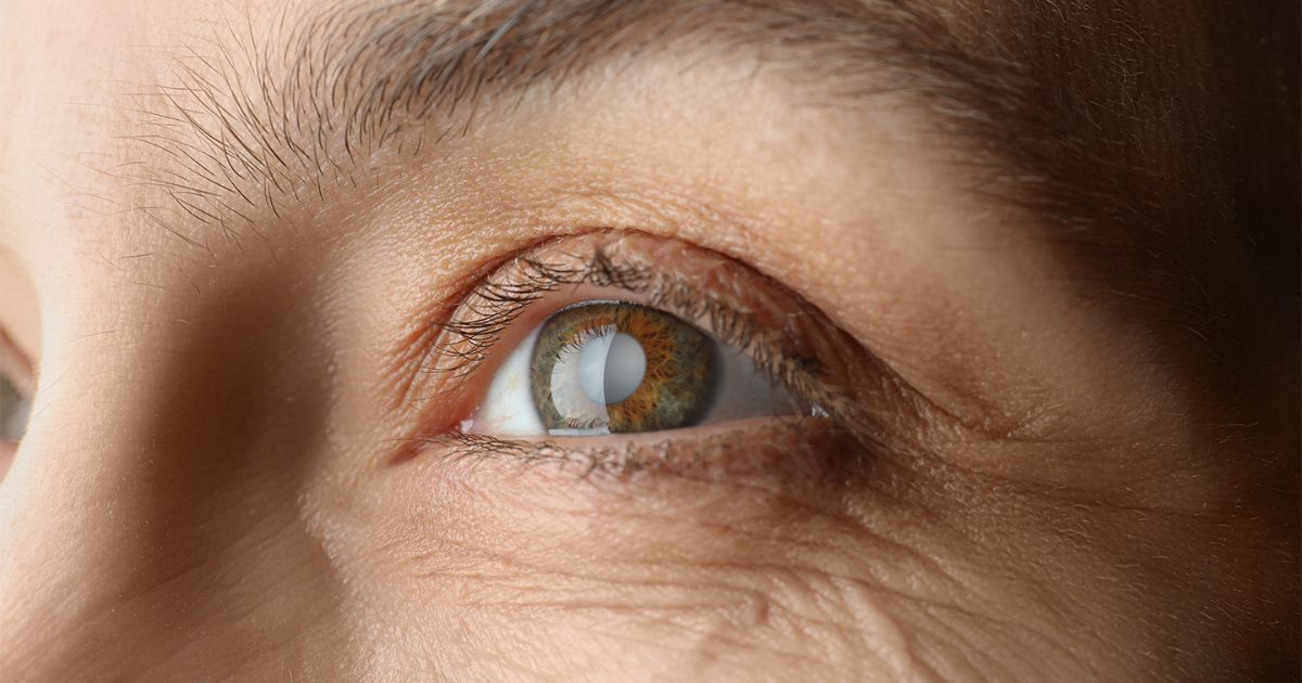
Cataracts are the leading cause of blindness worldwide, affecting more than 20 million people globally. The eye’s natural lens (crystalline lens) is responsible for focusing light to provide clear vision at various distances. When the lens becomes cloudy due to protein denaturation (commonly associated with aging, trauma, or prolonged corticosteroid use), light can no longer pass through properly, resulting in blurred vision resembling a foggy veil.

Illustration of vision affected by cataracts
Conjunctivitis
Conjunctivitis is inflammation of the transparent membrane covering the eyeball and inner eyelids. When caused by bacterial or viral infections, the condition can spread easily through contact with ocular secretions. However, it is generally not serious and does not cause permanent vision damage if diagnosed and treated promptly.
Digital Eye Strain
This condition is typically caused by prolonged and intensive visual focus on digital screens, combined with a reduced blinking rate that contributes to dry eye disease. Digital eye strain usually resolves quickly when the eyes are given adequate rest.
3. Serious Retinal and Optic Nerve Diseases
Glaucoma
Often referred to by ophthalmologists as the “silent thief of sight.” Glaucoma occurs when elevated intraocular pressure damages the optic nerve fibers. The disease gradually steals vision from the peripheral field toward the center, making early detection difficult. It is a leading cause of sudden or progressive vision loss that cannot be reversed once damage has occurred.
Retinal Detachment
Retinal detachment occurs when the retina separates from the underlying retinal pigment epithelium that nourishes it. Although usually painless, it can cause sudden vision loss and the appearance of a dark curtain or shadow across the visual field. This condition is a true ophthalmic emergency. If the retina is not promptly reattached, retinal cells may become deprived of oxygen, leading to irreversible damage and permanent vision loss.
Diabetic Retinopathy
A complication of diabetes that damages the tiny blood vessels within the retina, potentially leading to macular edema, vitreous hemorrhage, and severe vision impairment during working-age years.
4. Functional Vision Disorders and Other Causes
Color Blindness
Color blindness most commonly results from inherited abnormalities affecting the cone photoreceptor cells in the retina. Color perception is impaired when these pigments are defective or when cone cells respond abnormally to specific wavelengths of light.
Men have a significantly higher prevalence of color blindness (approximately 8%) compared to women (less than 1%) because the responsible gene is located on the X chromosome. Complete color blindness (seeing only black and white) is extremely rare. Most affected individuals are unable to distinguish certain colors, particularly red and green.
Night Blindness
A condition characterized by severely reduced vision or an inability to see in low-light environments or at night, often associated with damage to retinal rod cells or severe vitamin A deficiency.
Other Factors: Eye cancer (uveal melanoma), albinism (a deficiency of protective pigment that shields the eyes from UV radiation), ocular trauma, or intracranial injuries affecting the visual cortex in the occipital lobe.
Risk Factors and High-Risk Populations
According to the World Health Organization (WHO), factors that increase the risk of vision impairment include:
Table 1: Population Groups at High Risk of Vision Impairment
| Population Group | Primary Risk Factors | Common Associated Eye Conditions |
|---|---|---|
| Older Adults (Over 50 Years Old) | Natural aging processes affecting ocular cellular structures. | Cataracts, Age-Related Macular Degeneration (AMD), Presbyopia, Glaucoma. |
| Individuals with Chronic Diseases | Poorly controlled blood glucose levels and hypertension. | Diabetic Retinopathy, Retinal Artery or Vein Occlusion. |
| People with High Myopia (Greater than 6 Diopters) | Excessive elongation of the eyeball causing thinning of retinal and choroidal tissues. | Retinal Detachment, Myopic Chorioretinal Degeneration, Open-Angle Glaucoma. |
| Office Workers and Students | Prolonged near-work activities and intensive exposure to blue light. | Progressive Myopia, Computer Vision Syndrome, Asthenopia (Eye Strain). |
Warning Signs and Symptoms of Vision Impairment
Symptoms of vision impairment may develop gradually or occur suddenly, depending on the underlying cause.
Early and Common Symptoms
- Blurred vision, reduced sharpness, or difficulty seeing clearly at either near or far distances.
- Frequent eye strain and headaches, especially after prolonged reading or computer use.
- The need to squint or tilt the head to see objects more clearly.
- Double vision (diplopia), where a single object appears as two images.
- Difficulty adapting to dim lighting conditions or transitioning from bright to dark environments.
Urgent Warning Signs Requiring Immediate Medical Attention
If any of the following symptoms occur, patients should seek emergency ophthalmic care immediately, as they may indicate serious eye conditions capable of causing rapid and irreversible vision loss:
- Sudden vision loss or severe vision reduction in one or both eyes without pain.
- Severe eye pain accompanied by headache, nausea, and red eyes (typical symptoms of acute glaucoma).
- Persistent flashes of light or a dense appearance of floaters in front of the eyes.
- A sensation of a dark curtain blocking part or all of the field of vision.
- Sudden light sensitivity or seeing bluish-green halos around light sources.
Treatment and Management Methods for Vision Impairment
Depending on the specific cause, modern medicine offers effective medical, surgical, and visual-assistive treatment protocols.
Table 2: Comparison of Common Treatment Methods for Vision Impairment
| Cause Group | Treatment Method | Effectiveness | Limitations / Notes |
|---|---|---|---|
| Refractive Errors | Eyeglasses, Contact Lenses, Laser Surgery (LASIK, Femto-LASIK, ReLEx SMILE), Phakic Surgery (ICL). | Rapidly restores optimal vision and reduces dependence on glasses. | Laser procedures require adequate corneal thickness. |
| Cataracts | Artificial intraocular lens replacement surgery (Phaco or Laser Cataract Surgery). | Completely addresses the cause of blurred vision, with a success rate of over 95%. | Careful postoperative care is required to prevent infection. |
| Glaucoma | Intraocular pressure-lowering eye drops, SLT laser treatment, trabeculectomy. | Prevents disease progression and preserves remaining vision. | Cannot restore optic nerve damage that has already occurred. |
| Retinal Detachment | Scleral buckle surgery, vitrectomy, and gas/silicone oil tamponade. | Helps reattach the retina and salvage vision. | Visual recovery depends on how early or late intervention is performed. |
Visual Assistive Devices for People with Low Vision
For individuals with severe vision impairment that cannot be corrected by the medical treatments mentioned above, doctors may prescribe assistive devices to improve daily functioning:
- Telescopic lenses: Help users see distant objects such as license plates and signs.
- Handheld magnifiers or illuminated magnifiers: Help enlarge text when reading.
- Screen magnifiers and closed-circuit television (CCTV) systems: Electronic devices that magnify text onto a large, high-contrast screen.
Prevention and Protection of Visual Health
To maintain healthy eyes and minimize the risk of vision impairment, ophthalmology experts recommend the following habits:
Habits to Maintain
- Follow the 20-20-20 rule: When working on a computer, every 20 minutes, look at an object 20 feet away (approximately 6 meters) for at least 20 seconds to relax the ciliary muscles.
- Maintain an antioxidant-rich diet: Add foods rich in Vitamin A, C, E, zinc, lutein, and zeaxanthin, which are abundant in dark green vegetables such as kale and broccoli, fatty fish such as salmon, and berries.
- Protect your eyes from UV rays: Always wear sunglasses with UV protection (UV400) when going outdoors in sunny weather to help prevent early cataract formation.
- Ensure proper lighting for work: Always work and study in an environment with adequate standard lighting, and avoid reading or using a phone in the dark.
Things to Avoid
- Do not overwork your eyes continuously for long periods without rest.
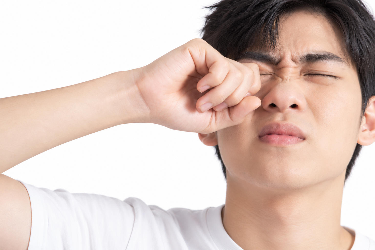
- Avoid the habit of rubbing your eyes with dirty hands, as this action may scratch the cornea and transmit bacteria that cause infection.
- Never self-purchase or use corticosteroid-containing eye drops without an ophthalmologist’s prescription, as these medications may increase the risk of cataracts and secondary glaucoma.
When Should You Have Regular Eye Examinations?
Eye examinations are not only for people who already have blurred vision. Proactive screening is the golden key to protecting your eyesight:
- Children: Should undergo at least one vision screening between the ages of 3 and 5 to detect refractive errors and amblyopia risk at an early stage.
- Adults aged 18–39 years: Should have a comprehensive eye examination every two years if no abnormal symptoms are present.
- Adults over 40 years old and individuals with underlying conditions (Diabetes, Hypertension): Should undergo eye examinations once a year to facilitate the early detection of cataracts, glaucoma, and retinal diseases.
Vision impairment can result from a wide range of causes, from common refractive errors that can be effectively managed with eyeglasses or modern refractive procedures to complex retinal diseases requiring urgent medical intervention. Establishing a routine of regular eye examinations today is one of the most effective ways to monitor, preserve, and protect your visual health for the long term.
 14/10/2026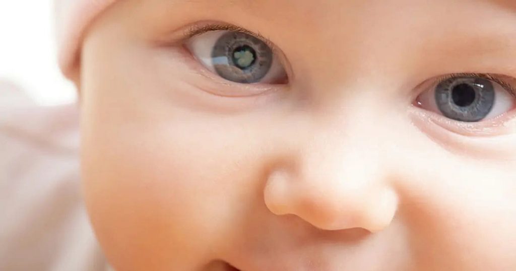










 0916.741.763
 Appointment