

 vi
vi 24-Sep-2026
 15,390
 Phakic EVO ICL involves implanting a lens inside the eye, where it is designed to remain long term. Because the lens must fit the anatomy of each individual eye, inappropriate sizing may increase the risk of unwanted complications. This article explains the science behind how the appropriate ICL size is selected for your eyes.

How is your ICL lens sized with micrometer-level precision?
Personalized ICL Sizing
Every eye has a unique anatomical structure. Ocular measurements vary from one person to another and may even differ between the right and left eyes of the same individual. Because the ICL is positioned just in front of the natural crystalline lens, within the narrow posterior chamber behind the iris, the size of the implant must closely match the anatomy of each eye.
One of the key goals for ophthalmic surgeons is to achieve an appropriate vault—the distance between the posterior surface of the ICL and the anterior surface of the natural crystalline lens. This space should be sufficient to keep the ICL safely separated from surrounding structures without excessive compression.
The Role of ANTERION in Phakic ICL Surgery
Before selecting an ICL size, the surgeon needs a detailed map of the internal structures of the eye. One technology used for this purpose is ANTERION, an anterior segment optical coherence tomography (Anterior Segment OCT) system.
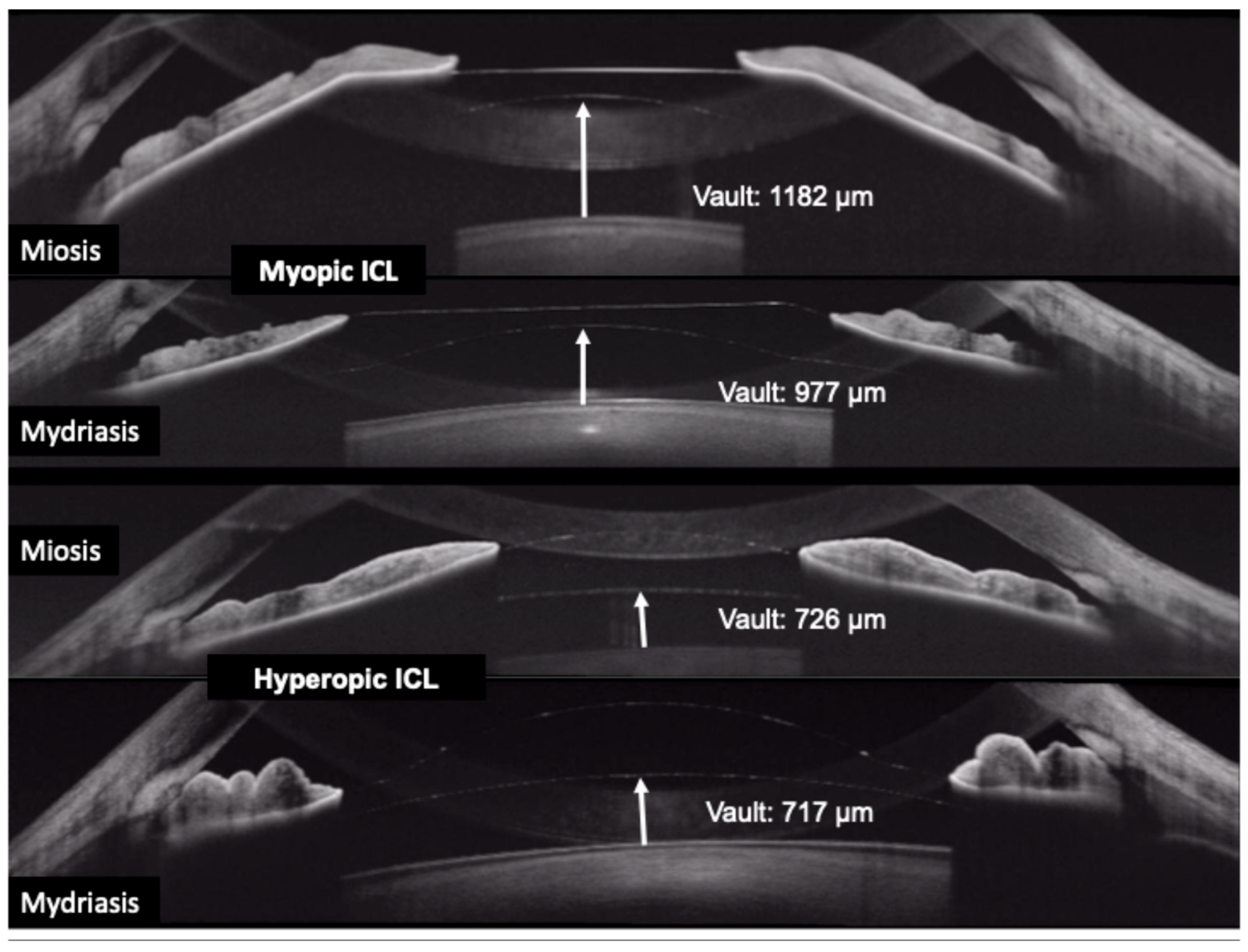
Anterior segment measurements obtained with ANTERION for Phakic ICL surgery
Unlike conventional measurement methods that may provide only a limited number of biometric parameters, ANTERION can capture high-resolution images of the entire anterior segment of the eye in a non-invasive, non-contact examination completed within seconds. These images allow the surgeon to assess measurements such as:
- Aqueous depth (AQD)—the fluid-filled distance in front of the natural crystalline lens
- Anterior chamber angle (ACA)—the angle formed between the cornea and iris, reflecting the available space within the anterior segment
- Crystalline lens thickness (LT)—an anatomical parameter that may influence the available space within the eye
- Additional measurements related to anterior chamber and angle anatomy
Together, these measurements form a highly individualized biometric profile of your eye.
Why Is ANTERION Used in Phakic ICL Planning?
The ANTERION system from Heidelberg Engineering can be incorporated into the preoperative assessment for Phakic ICL surgery because it provides detailed imaging and quantitative measurements of the anterior segment.

Why is ANTERION used in Phakic ICL planning?
High-resolution imaging: The system uses a long-wavelength 1300 nm swept-source OCT technology, enabling detailed visualization of structures throughout the anterior segment, from the cornea and iris to the crystalline lens.
Comprehensive measurements: ANTERION provides quantitative information on parameters such as anterior chamber depth, anterior chamber volume, and anatomical distances. After ICL implantation, the distance between the ICL and the natural crystalline lens is known as the vault, an important parameter in postoperative assessment and long-term lens positioning.
Supporting complication risk reduction: Detailed measurement of anterior segment anatomy can help surgeons select an appropriate ICL size and plan implantation more precisely. Appropriate sizing is important because an excessively high vault may contribute to angle narrowing or elevated intraocular pressure, whereas an excessively low vault may increase contact or proximity between the ICL and crystalline lens and potentially raise the risk of anterior subcapsular cataract formation.
What Does This Mean for You?
When ANTERION imaging is performed before ICL surgery, it is more than a routine diagnostic step. The surgeon is collecting detailed anatomical data that may help to:
- Select an ICL size that better matches the anatomy of your individual eye
- Reduce the risk of requiring lens exchange because of inappropriate sizing
- Optimize postoperative vault to maintain appropriate spacing between the ICL and the natural crystalline lens while preserving the surrounding ocular structures
In other words, the lens implanted in your eye is selected through a personalized process based on measurements that reflect the unique anatomy of your eyes.
Advice for Patients Considering ICL Surgery
If you are considering ICL surgery, you may want to ask your ophthalmologist which imaging and biometric technologies are used during preoperative assessment, how the ICL size is selected, and what postoperative vault is expected for your individual eyes. Understanding how these measurements contribute to ICL sizing can help you make a more informed decision about surgery.
Frequently Asked Questions About Phakic EVO ICL
1. Is ANTERION eye imaging painful or invasive?
No. ANTERION uses non-contact optical coherence tomography, does not require topical anesthesia, and typically takes only a few seconds to scan each eye.
2. Why can’t the same ICL sizing formula be used with every imaging device?
Different devices, such as ANTERION, Pentacam, and CASIA, use different optical technologies and image reconstruction algorithms. As a result, measurements obtained from one device may not be directly interchangeable with those from another.
3. What is ICL vault, and why is it important?
Vault is the distance between the posterior surface of the ICL and the anterior surface of the natural crystalline lens. An excessively high vault may contribute to angle narrowing and elevated intraocular pressure, while an excessively low vault may increase the risk of contact with the crystalline lens and anterior subcapsular cataract formation.
4. Which measurements have the greatest influence on ICL size selection?
Aqueous depth, anterior chamber angle measurements, and crystalline lens thickness are among the important anatomical parameters associated with postoperative ICL vault. These measurements are interpreted together with other biometric data when selecting the most appropriate lens size.
5. What happens if the wrong ICL size is selected?
In some cases, an inappropriately sized ICL may result in an excessively high or low vault and may require lens repositioning or exchange. This can involve additional surgical risk, recovery time, and cost, which is why precise preoperative measurement and sizing are essential.

 01/10/2026











 0916.741.763
 Appointment