

 vi
vi 30-Mar-2026
 30,590
 What Is Intraocular Pressure and Why Is It Important?
What Is Intraocular Pressure and Why Is It Important?
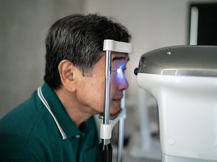
Intraocular pressure (IOP) is the pressure of the fluid inside the eye. It plays a critical role in maintaining the shape of the eyeball and ensuring that internal eye structures function properly.
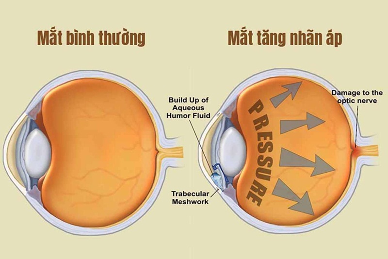
Under normal physiological conditions, the eye maintains a continuous circulation of aqueous humor: this fluid is produced behind the iris, flows forward through the anterior chamber, and is eventually drained out through the eye’s drainage system.
When the production and drainage of this fluid are balanced, intraocular pressure remains stable. However, if outflow is obstructed, pressure increases; conversely, if production decreases or leakage occurs, pressure may drop abnormally.
In healthy individuals, IOP typically ranges from 10–21 mmHg, and this is not just a number but a reflection of the eye’s physiological balance. When it falls outside this range, the risk of visual damage begins to emerge.
Classification of Intraocular Pressure
Classification of Intraocular Pressure
1. Normal Intraocular Pressure
At normal levels, intraocular pressure helps the eye maintain structural stability, ensures light is accurately focused onto the retina, and supports proper function of the optic nerve system. However, it is important to understand that “normal” is not the same for everyone, as some individuals may still develop optic nerve damage even when IOP is not elevated, a condition known as normal-tension glaucoma.
2. High Intraocular Pressure
When intraocular pressure remains elevated over time, the optic nerve is subjected to continuous compression and gradually becomes damaged. What makes this condition particularly concerning is that it often progresses without obvious symptoms in its early stages, yet the damage is irreversible once it occurs.
Clinical studies related to refractive procedures have reported that approximately 2.7% to 3.8% of cases may experience significant IOP elevation after intervention, and some of these may progress to glaucoma if not properly controlled. This highlights how even small disruptions in intraocular fluid balance can lead to long-term consequences.
3. Low Intraocular Pressure
Although less common, abnormally low intraocular pressure is not harmless. When IOP drops below physiological levels, particularly under 5 mmHg, the eye may lose structural stability, causing the retina to lose proper tension. This can result in blurred vision, increased risk of macular edema, and prolonged reduction in visual quality.
This condition is often associated with trauma, postoperative complications, or disorders affecting aqueous humor production.
Factors That Increase the Risk of IOP Disorders
Factors That Increase the Risk of IOP Disorders
Age
IOP disorders do not occur randomly but are often the result of cumulative risk factors over time. As people age, especially after 40–60 years, the eye’s drainage system becomes less efficient, leading to a gradual increase in the risk of elevated intraocular pressure.
Genetics
Genetic factors also play an important role. If there is a family history of glaucoma, your risk is significantly higher, even in the absence of early symptoms.
High Myopia
High myopia is not merely a refractive error; it is associated with structural changes in the eyeball that increase the risk of retinal damage and intraocular pressure imbalance.
Long-term Corticosteroid Use
Prolonged use of corticosteroids, whether in the form of eye drops, oral medication, or inhalation, can reduce aqueous humor outflow and lead to secondary glaucoma.
History of Eye Surgery or Trauma
Individuals who have undergone intraocular surgery or experienced eye trauma should be especially cautious, as these factors may cause long-term alterations in aqueous humor dynamics and affect intraocular pressure.
When Should You Check Intraocular Pressure?
Not everyone needs immediate IOP measurement, but certain warning signs should not be ignored. If your vision changes in ways that are difficult to explain, such as gradual loss of peripheral vision or a feeling that your visual field is no longer complete, this may indicate early optic nerve damage.
Seeing halos around lights, especially at night, is another important sign often associated with corneal edema or light scattering due to increased intraocular pressure.
In more severe cases, eye pain accompanied by nausea may indicate an acute glaucoma attack, which requires urgent medical attention. Similarly, red eyes combined with pain and vision loss warrant immediate IOP evaluation to rule out serious conditions.
Even in the absence of symptoms, individuals in high-risk groups, such as those over 40, with high myopia, a family history of glaucoma, or long-term steroid use, should undergo regular eye pressure checks.
IOP Measurement Is Not Just for Disease Detection
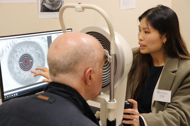
Many people assume that IOP should only be measured when problems arise. In reality, it is a crucial proactive monitoring tool. Regular check-ups help detect early changes before symptoms appear, track disease progression over time, and evaluate treatment effectiveness.
In glaucoma management, controlling intraocular pressure is the key factor in preserving remaining vision, as any damage that has already occurred is largely irreversible.
Treatment of Abnormal Intraocular Pressure
The goal of treatment is not merely to lower IOP values but to protect the optic nerve from progressive damage. In most cases, eye drops are used to reduce aqueous humor production or improve its outflow, forming the foundation of long-term management under medical supervision.
In certain situations, laser procedures may be applied to enhance fluid drainage. While effective, they may need to be repeated over time. When medication is insufficient to control IOP, surgical options are considered to create new drainage pathways or implant devices that facilitate fluid outflow.
Glaucoma does not cause immediate blindness; instead, it progresses slowly, silently, and cumulatively over time. Therefore, the timing of detection is crucial: early diagnosis can preserve vision, while late detection can only help limit further loss.
A simple eye pressure check today may be the key factor in protecting your vision for years to come.
 14/10/2026










 0916.741.763
 Appointment