

 vi
vi 21-Aug-2026
 23,465
 Retinal vein occlusion is one of the most common retinal vascular disorders and can cause sudden vision loss or gradually progressive visual impairment over time.
What is retinal vein occlusion?
Retinal vein occlusion is a condition in which a vein responsible for carrying blood away from the retina becomes blocked or has impaired blood flow. As a result, blood cannot drain normally from the retina, leading to increased pressure within the small blood vessels, retinal hemorrhage, and leakage of fluid into the surrounding tissues.
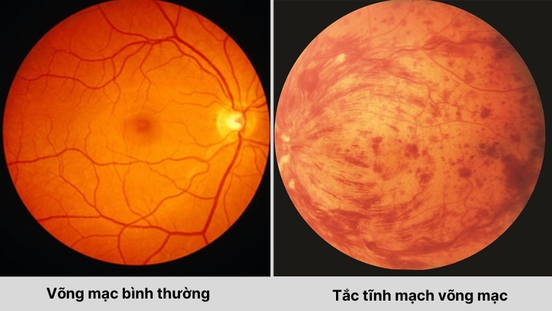
Normal retina and retinal vein occlusion
The retina is a layer of nerve tissue located at the back of the eye. Its primary function is to receive light and convert it into nerve signals that are transmitted to the brain. Within the retina, the macula is particularly important for central vision, enabling people to read, recognize faces, drive, and see fine details.
When the retinal venous system becomes blocked, the amount of oxygen reaching retinal cells may decrease. At the same time, damaged blood vessels can leak blood and fluid. If fluid accumulates in the macula, the patient may develop macular edema, one of the most common causes of vision loss in people with retinal vein occlusion.
In cases of severe retinal ischemia, abnormal new blood vessels known as neovascularization may also develop, increasing the risk of vitreous hemorrhage and neovascular glaucoma.
Is retinal vein occlusion dangerous?
The severity of retinal vein occlusion depends on the location of the blockage, the extent of the retina affected, the degree of retinal ischemia, and any associated complications.
Some mild cases may cause only moderate visual impairment. In contrast, severe occlusion, particularly when the central retinal vein is affected or when extensive retinal ischemia occurs, can lead to significant and prolonged vision loss.
Common types of retinal vein occlusion
Based on the location of the blocked blood vessel, retinal vein occlusion is divided into two main types.
1. Central retinal vein occlusion
Central retinal vein occlusion (CRVO) occurs when the central retinal vein, which drains blood from most of the retina, becomes blocked. Because a large portion of the retina may be affected, CRVO can cause widespread retinal hemorrhage and significant visual impairment.
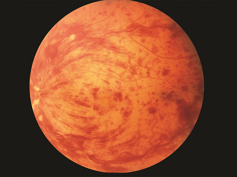
Central retinal vein occlusion
The condition usually affects one eye and may occur relatively suddenly. Depending on the degree of retinal ischemia, CRVO is generally classified into two groups:
- Non-ischemic CRVO: The retinal damage is generally less severe, and there is a better chance of preserving vision.
- Ischemic CRVO: A large area of the retina does not receive enough oxygen, resulting in a higher risk of severe vision loss and neovascularization.
People with hypertension, diabetes, glaucoma, dyslipidemia, or cardiovascular disease have a higher risk of developing CRVO.
2. Branch retinal vein occlusion
Branch retinal vein occlusion (BRVO) occurs when one of the smaller branches of the retinal venous system becomes blocked. This condition commonly develops at sites where a retinal artery and vein cross.
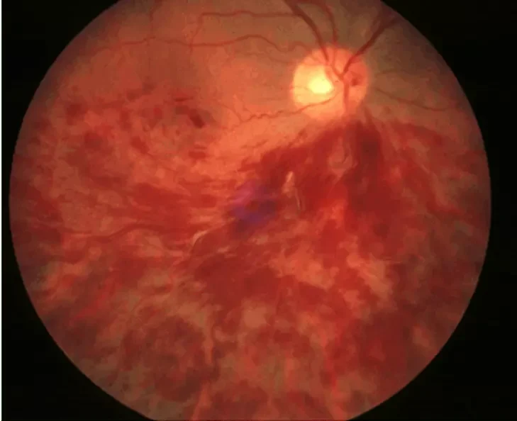
Branch retinal vein occlusion
When the arterial wall becomes hardened due to atherosclerosis, it may compress the adjacent vein, slowing blood flow and promoting the formation of a blood clot. BRVO usually affects a smaller area of the retina than CRVO. However, if the affected area involves the macula, the patient may still experience significant vision loss.
Causes of retinal vein occlusion
A single specific cause cannot always be identified. Retinal vein occlusion is often associated with a combination of changes in blood vessel walls, circulatory abnormalities, and systemic health conditions.
When blood flow through a retinal vein slows down or the vessel becomes narrowed, the risk of blood clot formation increases. The clot can obstruct blood flow and eventually lead to venous occlusion.
Atherosclerosis also plays an important role. Hardened retinal arteries may compress nearby veins at arteriovenous crossing points, reducing venous drainage and increasing the likelihood of blockage.
Who is at risk of retinal vein occlusion?
The risk of retinal vein occlusion increases with age and is generally higher among people with vascular or metabolic diseases.
Important risk factors include:
- Hypertension: One of the most important risk factors for retinal vein occlusion.
- Diabetes: Persistently high blood sugar levels can damage small blood vessels.
- Dyslipidemia: High cholesterol and triglyceride levels are associated with atherosclerosis.
- Atherosclerosis: Causes blood vessel walls to become thicker and harder.
- Glaucoma: Particularly associated with central retinal vein occlusion.
- Older age: The condition is more common in people over 50–60 years of age.
- Smoking: Damages blood vessels and increases the risk of cardiovascular disease.
- Obesity or physical inactivity: Indirectly increases the risk of hypertension, diabetes, and lipid disorders.
- Certain blood-clotting disorders: These may need to be considered, particularly in younger patients without common cardiovascular risk factors.
- A history of cardiovascular or vascular disease: This may be associated with underlying systemic circulatory problems.
Retinal vein occlusion most commonly affects only one eye. However, the other eye may also be affected in the future, particularly when systemic risk factors are not well controlled.
Signs and symptoms of retinal vein occlusion
A typical feature of retinal vein occlusion is painless vision loss, usually affecting one eye. The degree of visual impairment varies considerably between patients. Some people may notice only mild blurring, while others may experience a marked reduction in vision over a short period of time.
Common symptoms include:
- Blurred vision in one eye, with vision loss developing suddenly or progressively over several hours to several days.
- Distorted or warped vision and difficulty seeing fine details clearly.
- Dark areas or blurred spots in the visual field, as well as reduced central vision if the macula is affected.
- Black spots, floaters, or shadow-like areas if bleeding occurs into the vitreous.
Retinal vein occlusion is usually painless in its early stages. However, if the condition progresses to neovascular glaucoma, the patient may develop eye pain, redness, increased intraocular pressure, and severe visual impairment.
Treatment options for retinal vein occlusion
Intravitreal anti-VEGF injections
Anti-VEGF medications are currently among the most important treatments for macular edema caused by retinal vein occlusion. When the retina does not receive enough oxygen, levels of vascular endothelial growth factor (VEGF) can increase, making blood vessels more permeable and allowing fluid to leak into the retina.
Anti-VEGF medications help reduce blood vessel permeability, decrease macular edema, suppress abnormal new blood vessel growth, and improve or preserve vision in many patients. Some patients may require repeated injections and regular OCT monitoring according to their ophthalmologist’s recommendations.
Intravitreal corticosteroids
Corticosteroids can help reduce inflammation and decrease blood vessel permeability. This treatment may be considered in selected cases of macular edema, particularly when the response to other treatments is inadequate or when corticosteroid therapy is considered clinically appropriate.
Because corticosteroids can increase the risk of elevated intraocular pressure and cataract formation in some patients, regular monitoring of eye pressure is necessary.
Retinal laser photocoagulation
Laser treatment is not always used to manage macular edema now that modern intravitreal therapies have become widely available. However, retinal laser photocoagulation continues to play an important role when extensive retinal ischemia leads to neovascularization.
The purpose of laser treatment is to treat ischemic areas of the retina, thereby reducing the stimulus for abnormal blood vessel formation and lowering the risk of vitreous hemorrhage, iris neovascularization, and neovascular glaucoma. The specific treatment plan should be determined by a retinal specialist based on the type of retinal vein occlusion and the severity of retinal damage.
Can retinal vein occlusion recur?
An eye that has previously experienced retinal vein occlusion may continue to develop complications or experience another occlusive event in the future.
The other eye is not completely free from risk either, particularly in patients whose blood pressure, blood sugar, cholesterol levels, or cardiovascular conditions are not adequately controlled. Therefore, treatment of the eye should be combined with appropriate management of overall systemic health.
If sudden blurred vision, decreased vision, or loss of part of the visual field occurs in one eye, patients should seek prompt evaluation by an eye care specialist rather than waiting for the symptoms to resolve on their own.

 14/10/2026








 0916.741.763
 Appointment