

 vi
vi 12-Jul-2026
 36,385
Surface ablation refractive surgery, most notably photorefractive keratectomy (PRK), is a well-established and effective procedure for correcting refractive errors, including myopia, hyperopia, and astigmatism. However, one of the most clinically significant postoperative complications associated with PRK is the development of corneal haze.
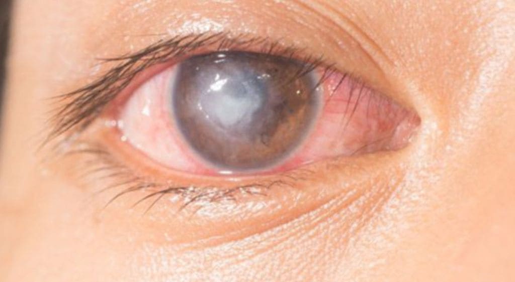
Corneal haze following surface ablation surgery
This article provides a comprehensive overview of postoperative corneal haze, covering its underlying biological mechanisms, major risk factors, clinical grading systems, and current evidence-based prevention and treatment strategies. The discussion is based on expert insights presented by Dr. Sharita Siregar at the Annual Congress of the European Society of Cataract and Refractive Surgeons (ESCRS) held in Copenhagen.
The Biological Mechanism Behind Corneal Haze Formation
Understanding why corneal haze develops requires an appreciation of the cornea’s natural wound-healing response. According to Dr. Siregar, removal of the corneal epithelium during PRK triggers a complex inflammatory cascade. Inflammatory mediators are rapidly released, stimulating epithelial proliferation while inducing apoptosis of stromal keratocytes.
Subsequently, transforming growth factor-beta (TGF-β) activates adjacent stromal keratocytes, driving their differentiation into myofibroblasts. Under normal physiological conditions, these myofibroblasts undergo apoptosis approximately 24 days after epithelial healing is complete. However, when this remodeling process becomes dysregulated and myofibroblasts persist within the anterior stroma, they produce excessive extracellular matrix and disorganized collagen, resulting in the formation of an opaque stromal layer that is clinically recognized as corneal haze.
Major Risk Factors
In many cases, postoperative corneal haze can be prevented by limiting PRK to patients with low-to-moderate refractive errors. Dr. Siregar strongly recommends: “To minimize the risk of corneal haze, PRK should generally be avoided in patients with high myopia, astigmatism greater than 3.0 D, or hyperopia exceeding +6.0 D. When treatment is unavoidable, intraoperative application of Mitomycin-C (MMC) should never be omitted.”
The primary risk factors associated with postoperative corneal haze include:
- High myopia or hyperopia greater than +6.0 D.
- Astigmatism exceeding 3.0 D.
- Irregular stromal bed following laser ablation.
- Delayed epithelial healing (e.g., epithelial basement membrane dystrophy).
- Severe dry eye disease.
- Excessive postoperative ultraviolet (UV) exposure.
- Failure to apply intraoperative Mitomycin-C (MMC).
Classification and Clinical Grading of Corneal Haze
In clinical practice, corneal haze is graded according to slit-lamp biomicroscopic findings using the Fantes grading scale, one of the most widely accepted classification systems.
| Grade | Slit-Lamp Clinical Findings |
|---|---|
| Grade 0 | The cornea is completely clear with no evidence of haze. |
| Grade 0.5 | Trace haze (faint reticular opacity) visible only with oblique slit-beam illumination. |
| Grade 1.0 | Mild haze without obscuring the fine details of the iris. |
| Grade 2.0 | Moderate haze with partial obscuration of iris details. |
| Grade 4.0 | Severe stromal opacity completely obscuring the iris and pupil, resulting in significant visual impairment. |
Corneal haze can also be classified according to the timing of onset:
- Early-onset haze: The most common presentation, typically developing within the first three months after surgery. In most patients, the haze gradually resolves spontaneously within one year.
- Late-onset haze: Develops more than three months after surgery and may persist for up to three years. This form is more concerning because it can lead to significant visual deterioration, permanent corneal scarring, and persistent visual disturbances.
Clinical research findings: Dr. Siregar is currently conducting a prospective study involving 173 eyes from 139 patients who underwent PRK for myopia correction. Preliminary findings demonstrate that both early- and late-onset corneal haze occur significantly more frequently in eyes treated for myopia greater than −6.00 D compared with eyes having lower refractive errors.
Interestingly, the study also found no statistically significant difference in haze incidence between short-duration MMC exposure (20–60 seconds) and longer application times (60–120 seconds).
Medical Management with Topical Corticosteroids
Most cases of postoperative corneal haze respond favorably to topical corticosteroid therapy. Dr. Siregar recommends the following treatment protocols:
- Early-onset haze: Instill Prednisolone acetate 1% every four hours. Patients should be re-evaluated at two weeks, with corticosteroid therapy continued for a total duration of approximately 12 weeks.
- Late-onset haze: Increase dosing frequency of Prednisolone acetate 1% to every two to three hours during the first four weeks, with close follow-up examinations scheduled at weeks 2 and 4.
Surgical Management for Persistent Corneal Haze
In the small proportion of patients who fail to respond adequately to medical therapy, surgical intervention should be considered based on the depth of stromal involvement.
Superficial haze (less than 15 μm): Mechanical epithelial debridement followed by intraoperative MMC application and postoperative Prednisolone acetate 1% every four hours is generally recommended.
Deep stromal haze (greater than 15 μm): Phototherapeutic keratectomy (PTK) or repeat PRK combined with 0.02% Mitomycin-C represents the preferred surgical approach. Postoperatively, patients should receive a bandage contact lens, topical non-steroidal anti-inflammatory drugs (NSAIDs), preservative-free artificial tears, and a structured 12-week topical corticosteroid regimen.
To optimize corneal healing and reduce the risk of recurrence, patients are advised to wear UV-blocking sunglasses whenever outdoors and consider oral supplementation with vitamin C and vitamin D to support collagen synthesis and corneal tissue remodeling.
Reference: Based on the scientific presentation by Dr. Sharita Siregar at the ESCRS Annual Congress 2025, Copenhagen.

 14/10/2026









 0916.741.763
 Appointment