

 vi
vi 16-Jul-2026
 28,880
Why Is a Retinal Examination Essential Before Refractive Surgery?
The Higher Your Myopia, the Greater the Risk of Retinal Damage
To understand why, it is important to first look at the anatomy of a myopic eye. In people with normal vision, the eyeball is nearly spherical. However, in individuals with myopia—particularly axial myopia—the eyeball becomes abnormally elongated from front to back.
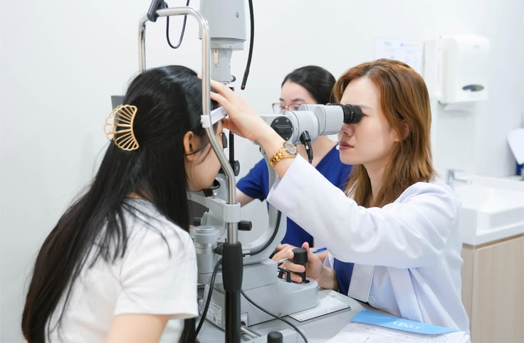
Why a Retinal Examination Before Vision Correction Surgery Is More Important Than You Think
As the eyeball elongates, the internal structures of the eye—especially the retina, the light-sensitive tissue lining the back of the eye—become stretched, much like an overinflated balloon. Over time, this mechanical stretching causes the retina to become thinner, weaker, and more susceptible to degeneration, particularly in the peripheral retina. As a result, the risks of the following conditions increase significantly:
- Peripheral retinal degeneration: Degenerative changes such as lattice degeneration and snail-track degeneration weaken the retinal structure.
- Retinal holes and retinal tears: Small breaks or holes can develop when the retina is subjected to excessive traction.
- Retinal detachment: The most serious complication, occurring when vitreous fluid passes through a retinal tear and separates the retina from the underlying tissue that nourishes it, potentially leading to permanent vision loss.
Vision Correction Surgery Does Not Treat Retinal Disease
This is one of the most common misconceptions among patients. Modern refractive procedures such as LASIK, SMILE Pro, and Phakic ICL surgery are designed to correct refractive errors. LASIK and SMILE Pro reshape the cornea using laser technology, while Phakic ICL involves implanting a lens inside the eye.
All of these procedures correct refractive errors only. They enable light to focus accurately on the retina so you can see clearly without glasses or contact lenses. However, they do not shorten the elongated eyeball or restore retinal thickness.
In other words, even after successful vision correction surgery, although you may no longer need glasses, the internal structure of your eye remains that of a myopic eye. Any underlying retinal abnormalities will still be present unless they are detected and treated beforehand.
Serious Retinal Conditions That Can Be Detected During a Dilated Retinal Examination
A comprehensive retinal examination allows ophthalmologists to detect retinal abnormalities while they are still in the early stages, such as small degenerative lesions or retinal breaks. Early diagnosis enables timely laser treatment and provides the best opportunity to protect long-term vision.
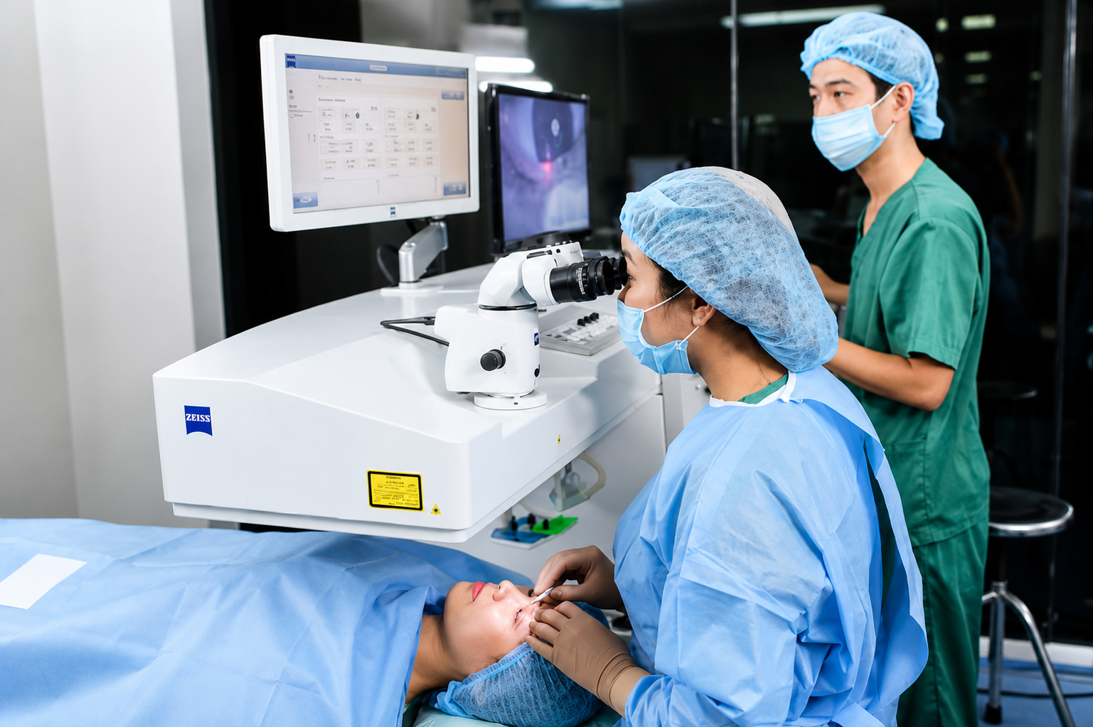 Serious Retinal Diseases That Can Be Detected During a Retinal Examination[/caption>
Peripheral Retinal Degeneration
- What is it? Peripheral retinal degeneration refers to thinning and structural changes in the outer retina, including conditions such as lattice degeneration and snail-track degeneration.
- Who is at higher risk? Individuals with myopia greater than -3.00 diopters or an axial length exceeding 25 mm.
- Does it require treatment before surgery? Your ophthalmologist will evaluate the severity of the lesion. If there is a significant risk of progression to a retinal tear, preventive laser photocoagulation is recommended before refractive surgery.
Retinal Holes
- Why do they occur? Retinal holes may develop when an area of the retina becomes extremely thin or when vitreous traction creates a small opening in the retina.
- Symptoms: In most cases, retinal holes produce no obvious symptoms during the early stages, and vision often remains normal.
- When is preventive laser treatment necessary? If the retinal hole is considered at high risk for retinal detachment, laser photocoagulation is performed around the hole to create scar tissue that seals the surrounding retina.
Retinal Tears
- Cause: Retinal tears typically occur when sudden vitreous traction pulls on areas of retina already weakened by high myopia or following ocular trauma.
- How serious is it? A retinal tear is a medical emergency because it significantly increases the risk of retinal detachment.
- Why should it be treated before refractive surgery? Laser photocoagulation seals the retinal tear before surgery, helping prevent retinal detachment and protecting the retina during and after the refractive procedure.
Retinal Detachment
When the retina separates completely from the underlying choroid that supplies it with oxygen and nutrients, retinal cells begin to die rapidly. Without prompt surgical treatment, permanent vision loss may occur.
Warning signs include:
- Frequent flashes of light, especially in the peripheral vision or in dark environments.
- A sudden increase in floaters, often appearing as numerous dark spots or cobweb-like shadows.
- A dark curtain or shadow blocking part of the visual field.
Can You Still Have Vision Correction Surgery If a Retinal Problem Is Found?
Many patients become anxious after learning they have a retinal abnormality, fearing they are no longer candidates for refractive surgery. In reality, modern ophthalmology offers clear management strategies depending on the specific condition.
- Scenario 1: No retinal abnormalities (or only stable, non-progressive lesions)
You can safely proceed with the standard refractive surgery process.
- Scenario 2: High-risk peripheral retinal degeneration or retinal holes/tears
Your ophthalmologist will recommend preventive retinal laser photocoagulation. This is a minimally invasive outpatient procedure that typically takes only 5–10 minutes. The laser creates a protective barrier of scar tissue around the lesion. After treatment, patients generally wait one to two weeks for the retina to heal before proceeding with refractive surgery.
- Scenario 3: Active retinal detachment or severe retinal disease (such as retinal hemorrhage or inflammation)
In these situations, preserving vision becomes the top priority. Refractive surgery must be postponed while specialized retinal treatment—such as scleral buckling or vitrectomy—is performed. Once the retina has stabilized, often several months later, your ophthalmologist can reassess whether vision correction surgery is safe and appropriate.
Who Requires a More Comprehensive Retinal Examination?
Every patient considering refractive surgery should undergo a retinal examination. However, if you belong to one of the following groups, your retina should be evaluated even more carefully due to the higher risk of retinal complications:
- People with high myopia (greater than -6.00 diopters) or rapidly progressing myopia.
- Individuals with an abnormally long axial eye length (greater than 25.5 mm).
- People experiencing numerous floaters or episodes of flashing lights in their peripheral vision.
- Individuals with a history of significant eye or head trauma resulting from accidents or contact sports.
- Those with a family history of retinal detachment in first-degree relatives.
- Patients with systemic conditions that affect the retinal blood vessels, such as diabetes or hypertension.
- Individuals who have previously undergone eye surgery or other major ophthalmic procedures.
How to Protect Your Retina After Vision Correction Surgery
Caring for your eyes does not end after the first few weeks of using postoperative eye drops. Maintaining long-term retinal health is essential for preserving clear vision for years to come. Here are several recommendations:
- Attend all scheduled follow-up visits, typically one day, one week, one month, three months, and six months after surgery.
- Continue having comprehensive eye examinations, including a dilated retinal examination at least once a year if you previously had high myopia, even if your vision remains excellent.
- Protect your eyes and head from injury. Avoid activities with a high risk of impact, such as boxing, martial arts, or highly competitive contact sports.
- Keep systemic conditions such as diabetes and hypertension well controlled to reduce the risk of retinal vascular complications.
- Pay close attention to any changes in your vision, and seek immediate ophthalmic care if you notice unusual symptoms such as flashes, floaters, or sudden vision loss.

 01/10/2026












 0916.741.763
 Appointment