

 vi
vi 18-Jul-2026
 46,645
 What Is Retinal Detachment?
The retina is a thin layer of nerve tissue located at the back of the eye. It functions much like the film in a camera—capturing light, converting it into nerve signals, and transmitting those signals to the brain, allowing us to see images.
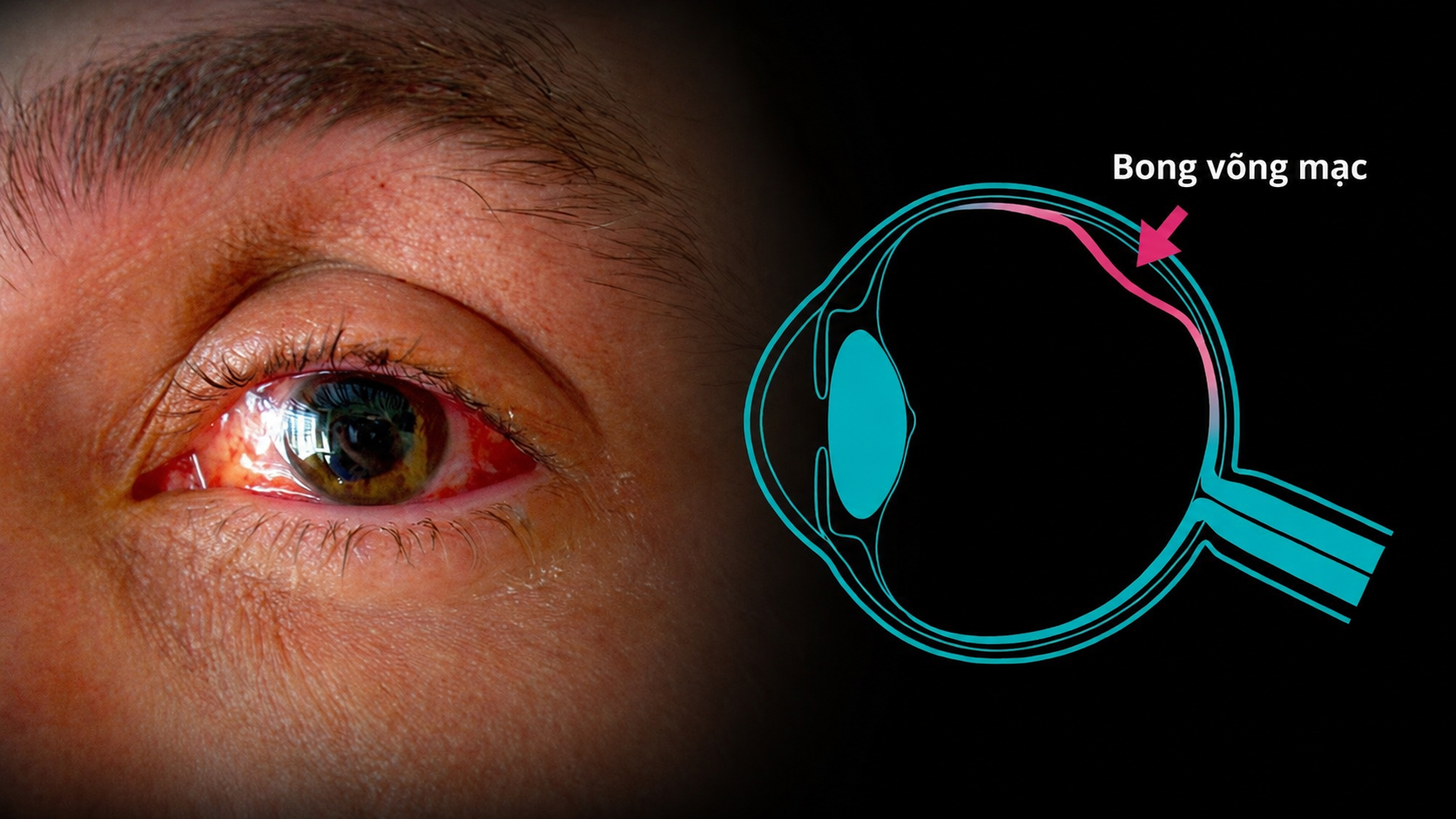
Photo of a patient with retinal detachment
Retinal detachment occurs when the neurosensory retina separates from the underlying retinal pigment epithelium (RPE) and the blood vessels that nourish it. Once deprived of its blood supply, retinal nerve cells can rapidly become damaged and die. Without prompt diagnosis and surgical treatment, retinal detachment may result in permanent vision loss or blindness.
Why Does High Myopia Increase the Risk of Retinal Detachment?
From an anatomical perspective, high myopia (typically defined as -6.00 diopters or greater) occurs when the axial length of the eye (the distance from the front to the back of the eyeball) becomes abnormally elongated.
The retina is the light-sensitive nerve tissue lining the back of the eye, responsible for capturing visual information and transmitting it to the brain. As the eyeball elongates, the retina is stretched excessively and gradually becomes thinner over time. This thinning weakens the retinal tissue, making it more susceptible to degeneration, retinal tears, and ultimately detachment from the underlying nourishing tissue, which can lead to permanent vision loss if not treated promptly.
To better understand this relationship, below are three key mechanisms explaining why high myopia increases the risk of retinal detachment:
1. Retinal Stretching
Imagine the retina as the rubber surface of a balloon. When a balloon is inflated too much (similar to an elongated eyeball in high myopia), the rubber becomes extremely thin and much easier to tear.

Why does high myopia increase the risk of retinal detachment?
In highly myopic eyes, the retina is subjected to continuous stretching. Over time, this reduces the natural elasticity of the retinal tissue, creating thin, fragile areas that are prone to developing retinal tears.
2. Peripheral Retinal Degeneration
As the eyeball enlarges, it becomes more difficult for blood and nutrients to adequately reach the far peripheral regions of the retina. As a result, people with high myopia are more likely to develop peripheral retinal degeneration (such as lattice degeneration and other peripheral degenerative changes).
In these degenerated areas, the retinal structure becomes severely weakened, making it extremely thin and fragile. Even minor mechanical forces or internal vitreous traction can cause small retinal breaks to develop.
3. Vitreous Changes (Mechanical Traction)
The inside of the eye is filled with the vitreous humor, a transparent gel that helps maintain the eye’s shape. In people with high myopia, the vitreous tends to liquefy much earlier than in individuals with normal vision.
As the vitreous liquefies, the remaining gel begins to shrink and move more freely within the eye. During this process, the vitreous may adhere to the already weakened retina and exert significant traction, causing retinal tears. Fluid can then pass through these tears, separating the retina completely from the back of the eye.
Early Warning Signs of Retinal Detachment
Retinal detachment is an ophthalmic emergency. If you have high myopia, pay close attention to your eye health and seek immediate medical attention if you experience any of the following symptoms:
- Flashes of light: Sudden flashes or streaks of light appearing in your vision (similar to camera flashes or small lightning bolts), especially in your peripheral vision. This may indicate that the vitreous is pulling on the retina.
- A sudden increase in floaters: A rapid appearance of numerous black spots, cobweb-like strands, threads, or floating specks drifting across your field of vision.
- A “curtain” over your vision: A dark shadow or curtain gradually covering part or all of your visual field, as though a blind is being pulled across your vision.
Expert recommendation: Individuals with high myopia should undergo a comprehensive eye examination every six months, including a dilated fundus examination, to detect and treat retinal degeneration or tears before retinal detachment develops.

 01/10/2026












 0916.741.763
 Appointment