

 vi
vi 23-Aug-2026
 23,465
 Diabetic macular edema is one of the serious vision-threatening complications in people with diabetes.
What is diabetic macular edema?
Diabetic macular edema (DME) is a condition in which fluid accumulates in the macula due to abnormal changes in the retinal blood vessels associated with diabetes.
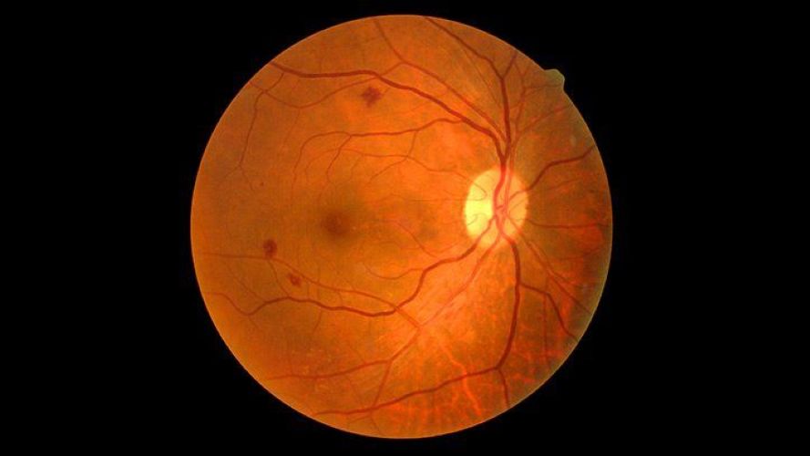
Diabetic macular edema: Causes, symptoms, complications, and treatment
At the center of the retina is a small area called the macula. This area is primarily responsible for central vision, facial recognition, reading, seeing fine details, and distinguishing colors. In people who have had diabetes for a long time, persistently high blood glucose levels can damage the walls of the retinal capillaries. These blood vessels may become weakened and more permeable, allowing fluid, lipids, and other blood components to leak into the retinal tissue.
When this leakage occurs in or near the macula, fluid accumulates and causes the area to thicken and swell. This condition is known as diabetic macular edema. DME may affect one or both eyes. The severity of damage does not necessarily develop equally in both eyes, so some patients may still feel that their vision is relatively normal if the other eye has not yet been significantly affected.
The relationship between diabetic retinopathy and macular edema
Macular edema is closely associated with diabetic retinopathy, a common eye complication of both type 1 and type 2 diabetes.
When blood glucose remains elevated over a prolonged period, the small blood vessels in the retina may become damaged. Some capillaries may dilate, become blocked, or leak, while in more advanced stages, abnormal and fragile new blood vessels may develop on the retina.
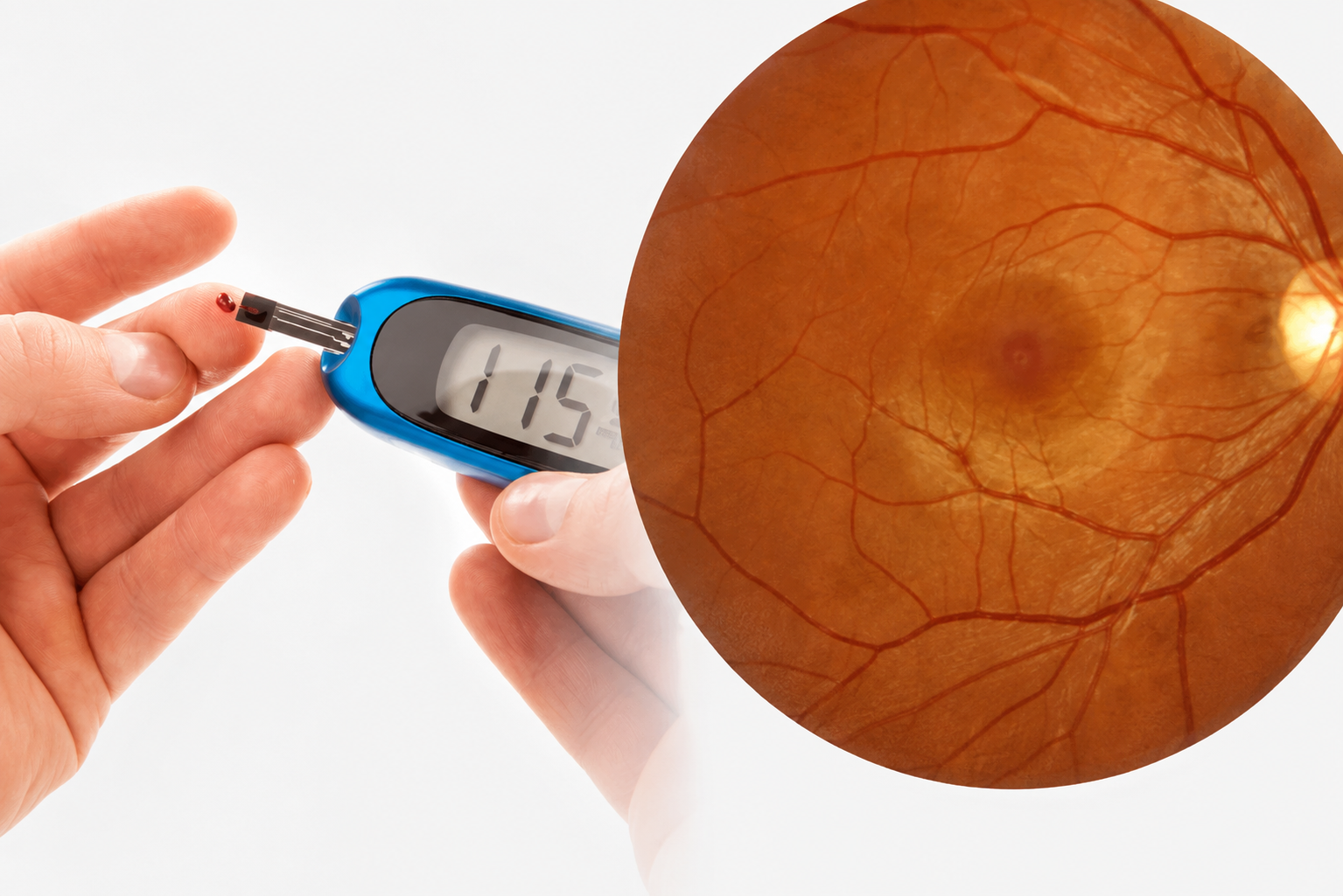
The relationship between diabetic retinopathy and macular edema
Macular edema can occur at different stages of diabetic retinopathy and does not necessarily develop only after the disease has progressed to the proliferative stage.
It is also one of the major causes of visual impairment in people with diabetes. If the damage persists, photoreceptor cells and neural structures in the macula may be affected, making visual recovery more limited.
Causes of diabetic macular edema
The underlying cause of macular edema is damage to the retinal microvasculature due to diabetes. When blood glucose remains elevated for a long period, the endothelial cells lining the blood vessels may become damaged, while the blood-retinal barrier becomes less stable. This increases capillary permeability and allows fluid to leak into the retinal tissue.
In addition, reduced oxygen supply in certain areas of the retina may stimulate the body to produce higher levels of vascular endothelial growth factor, commonly known as VEGF. VEGF increases blood vessel permeability and contributes to the development of abnormal blood vessels. Therefore, it is one of the key mechanisms involved in the development and progression of diabetic macular edema.
Who is at higher risk of developing macular edema?
Not everyone with diabetes will develop macular edema. However, the risk tends to increase when diabetes has been present for many years or is poorly controlled.

Normal eye and eye with diabetic macular edema
Several factors may increase the risk, including:
- Frequently elevated blood glucose levels or HbA1c that is not adequately controlled.
- A long duration of diabetes.
- Existing diabetic retinopathy.
- High blood pressure that is not well controlled.
- Dyslipidemia or high cholesterol levels.
- Diabetes-related kidney disease.
- Being overweight or obese and having a sedentary lifestyle.
- Smoking.
- Certain hormonal changes or systemic health conditions that may accelerate the progression of diabetic retinopathy.
The more risk factors a patient has, the more important it is to undergo regular eye examinations.
Signs and symptoms of diabetic macular edema
One of the challenges of macular edema is that it may progress for some time without causing obvious symptoms. When swelling begins to affect the central part of the macula, patients may notice changes such as:
Blurred central vision
This is a common symptom. Patients may notice that images are no longer sharp, particularly when reading small print, looking at a smartphone screen, or viewing another person’s face.
Distorted vision
Straight lines, such as door frames, table edges, or lines of text, may appear bent, wavy, or distorted.
Dark areas in the center of vision
Some people may notice a dark spot, blurred patch, or blank area in the center of the image they are looking at.
Difficulty distinguishing colors
Colors may appear faded, less vivid, less contrasted, or more difficult to distinguish than before.
Fluctuating vision
Vision may vary at different times, particularly when blood glucose levels fluctuate significantly.
If a person with diabetes notices sudden vision loss, distorted images, or a new dark area in the field of vision, they should visit an ophthalmology clinic for examination rather than waiting for the symptoms to improve on their own.
Is diabetic macular edema dangerous?
The severity of diabetic macular edema depends on the location and extent of swelling, how long the condition has been present, and the structural condition of the macula.
If the swelling persists, nerve cells and photoreceptor cells in the macula may become damaged. This can reduce the potential for visual recovery even after the accumulated fluid has been brought under control.
The condition may lead to consequences such as:
- Reduced central vision.
- Difficulty reading books or using smartphones and computers.
- Difficulty recognizing faces.
- Reduced ability to drive safely.
- Difficulty performing work that requires precise vision.
- Reduced ability to perform self-care and live independently when vision loss becomes severe.
Macular edema generally does not cause complete loss of peripheral vision in the same way that some other eye diseases may, but severe damage to central vision can still have a major impact on quality of life.
How can diabetic macular edema be prevented?
Not every case can be completely prevented, but the risk can be significantly reduced through proper diabetes management and regular eye examinations. Patients should maintain an appropriate diet, exercise as recommended, take medications as prescribed, and regularly monitor relevant metabolic indicators.
Most importantly, patients should not wait until their vision becomes blurred before having an eye examination. Diabetic retinopathy can develop before causing any noticeable symptoms. Regular dilated retinal examinations allow ophthalmologists to detect early microvascular changes and determine the appropriate timing for monitoring or treatment.
The recommended frequency of eye examinations depends on the type of diabetes, how long the patient has had the disease, the severity of retinal damage, pregnancy status, and overall health. People who already have diabetic retinopathy or macular edema generally require more frequent follow-up examinations.

 01/10/2026













 0916.741.763
 Appointment