

 vi
vi 23-Mar-2026
 37,050
 A comprehensive, standardized eye examination process can help detect risk factors early, personalize treatment methods, and optimize visual outcomes after surgery.
What is a comprehensive pre-refractive surgery exam?

What is a comprehensive pre-refractive surgery exam?
A comprehensive eye exam is a process of thoroughly evaluating key eye parameters using advanced diagnostic equipment, combined with clinical assessment by an ophthalmologist.
Unlike a standard vision check, a comprehensive exam focuses on:
- Accurate evaluation of refractive errors
- Assessment of corneal and retinal structures
- Analysis of visual quality under different conditions
- Detection of underlying conditions that may affect surgery
Therefore, this is a mandatory step before deciding on any vision correction procedure.
10 essential steps in a comprehensive eye exam before vision correction
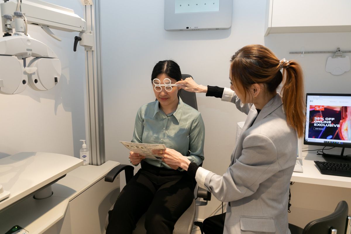
10 essential steps in a comprehensive eye exam before vision correction
1. First refraction test: Determine myopia, astigmatism, and hyperopia
This step aims to accurately determine the current level of refractive error, as each eye may differ. If the initial measurement is incorrect, the entire treatment plan may be affected. It provides the foundation for doctors to know how much correction is needed, avoiding undercorrection or overcorrection after surgery.
2. Second refraction test: Evaluate actual visual performance
Beyond numerical measurements, doctors will assess:
- Subjective vision (patient perception)
- Objective vision (measured by devices)
This combination helps eliminate errors caused by instruments or eye accommodation, ensuring an accurate assessment of real visual quality and preventing treatment inaccuracies.
3. 2D corneal topography: Detect keratoconus
The main purpose is to rule out keratoconus or other corneal structural abnormalities. This is critical because performing laser surgery on an unstable cornea may lead to serious complications and irreversible vision loss. In other words, this step determines whether you are eligible for surgery.
4. Corneal thickness measurement
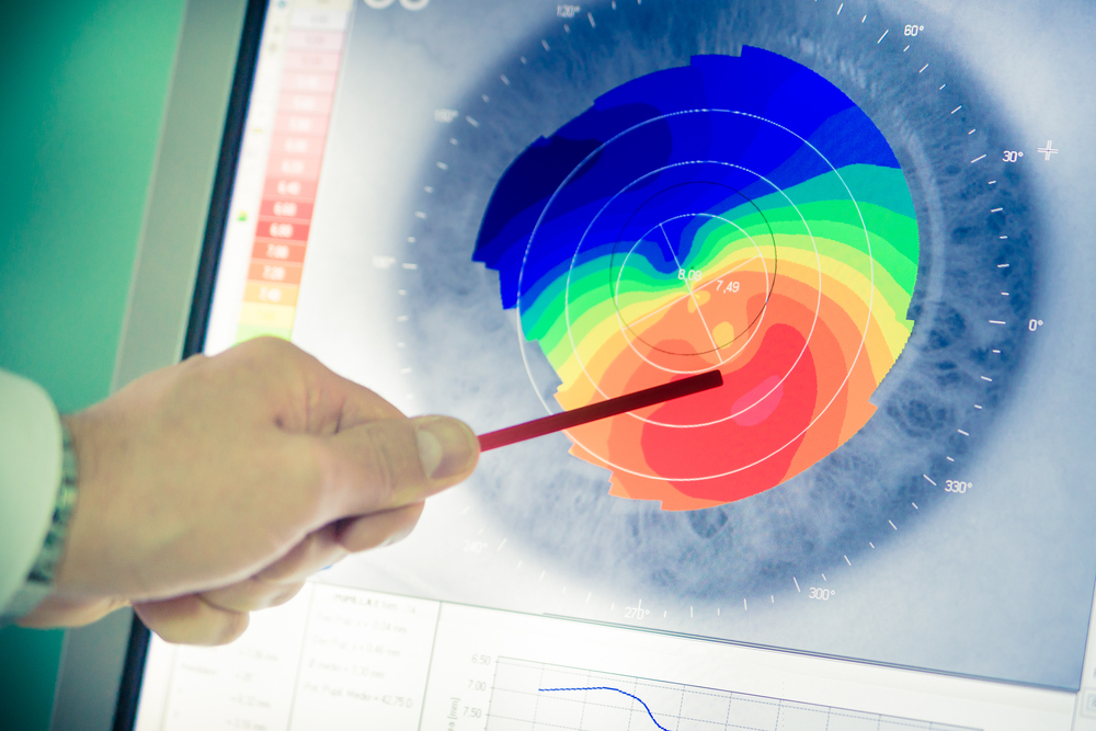
Corneal thickness measurement helps determine the appropriate treatment method
Each vision correction method requires a minimum corneal thickness. If the cornea is too thin, surgery may lead to long-term structural instability. This step helps doctors decide whether laser surgery is suitable or if alternative options such as Phakic ICL should be considered to preserve corneal integrity.
5. Corneal diameter measurement
This parameter determines the appropriate optical zone size, ensuring light entering the eye is properly processed. If the treatment zone is too small, patients may experience halos or glare at night. Measuring corneal diameter helps minimize these visual disturbances.
6. Corneal curvature measurement
Since the cornea plays a key role in focusing light, measuring its curvature helps determine whether light is evenly focused. Irregularities such as uneven astigmatism may require adjustments or reconsideration of surgery. Skipping this step may result in suboptimal visual outcomes.
7. Pupil size measurement
A larger pupil, especially in low-light conditions, allows more light into the eye. Without proper measurement, patients may experience glare or halos after surgery, particularly when driving at night. This step helps doctors choose the most suitable method to minimize these issues.
8. Fundus examination
The goal of this step is to examine the retina, which ultimately determines visual function. After pupil dilation, doctors will assess the entire fundus to detect:
- Retinal degeneration
- Retinal tears
- Other underlying conditions
If present, early intervention is required to prevent serious complications after surgery.
9. Personalized 3D corneal mapping
This advanced step uses high-resolution scanning technology to map up to 150,000 points of the eye, creating a detailed 3D model.
Through this, doctors can:
- Analyze higher-order aberrations
- Evaluate vision in low-light conditions
- Design a personalized treatment plan
This is a key factor in optimizing visual quality after surgery.
10. One-on-one consultation with a doctor
After completing all tests, this consultation integrates all collected data to determine the most suitable treatment for each eye. It is also when patients gain a clear understanding of the benefits, risks, and realistic expectations before making a decision.
Why should you not skip a comprehensive exam?
Many people want quick vision correction and tend to skip or rush through the examination process. However, this can lead to serious risks:
- Choosing the wrong treatment method
- Increased risk of complications
- Suboptimal visual outcomes
On the other hand, a thorough examination process helps:
| Key Parameters | Clinical Significance |
|---|---|
| Accurate refraction (error < 0.25D) | Prevents under- or over-correction after surgery |
| Corneal thickness (typically ≥ 500µm) | Determines eligibility for laser procedures |
| Corneal curvature & topography | Detects early keratoconus and structural abnormalities |
| Pupil size in low light | Predicts risk of glare and halos at night |
| Retinal condition | Prevents retinal tears or detachment post-surgery |
| Higher-order aberrations (HOAs) | Directly impacts night vision quality |
| 3D corneal mapping (100,000+ data points) | Enables fully personalized treatment planning |
| Comprehensive doctor evaluation | Ensures correct method and proper indication |
In reality, the comprehensive exam is when doctors fully “read” your eyes—from structure to visual function. Every parameter plays an important role in building a safe and effective treatment plan.
If you are considering vision correction, do not rush. Start with a thorough eye exam to ensure your decision is well-informed and delivers clear, long-lasting vision for years to come.

 01/10/2026











 0916.741.763
 Appointment