

 vi
vi 22-May-2026
 78,850
 Overview
In Phakic ICL surgery, one of the factors that determines long-term safety and visual quality lies in extremely small parameters: ICL curvature, lens size, ICL vault, and the way the lens interacts with ocular structures.
What Is ICL Curvature?

ICL Curvature and Its Interaction with Ocular Structures
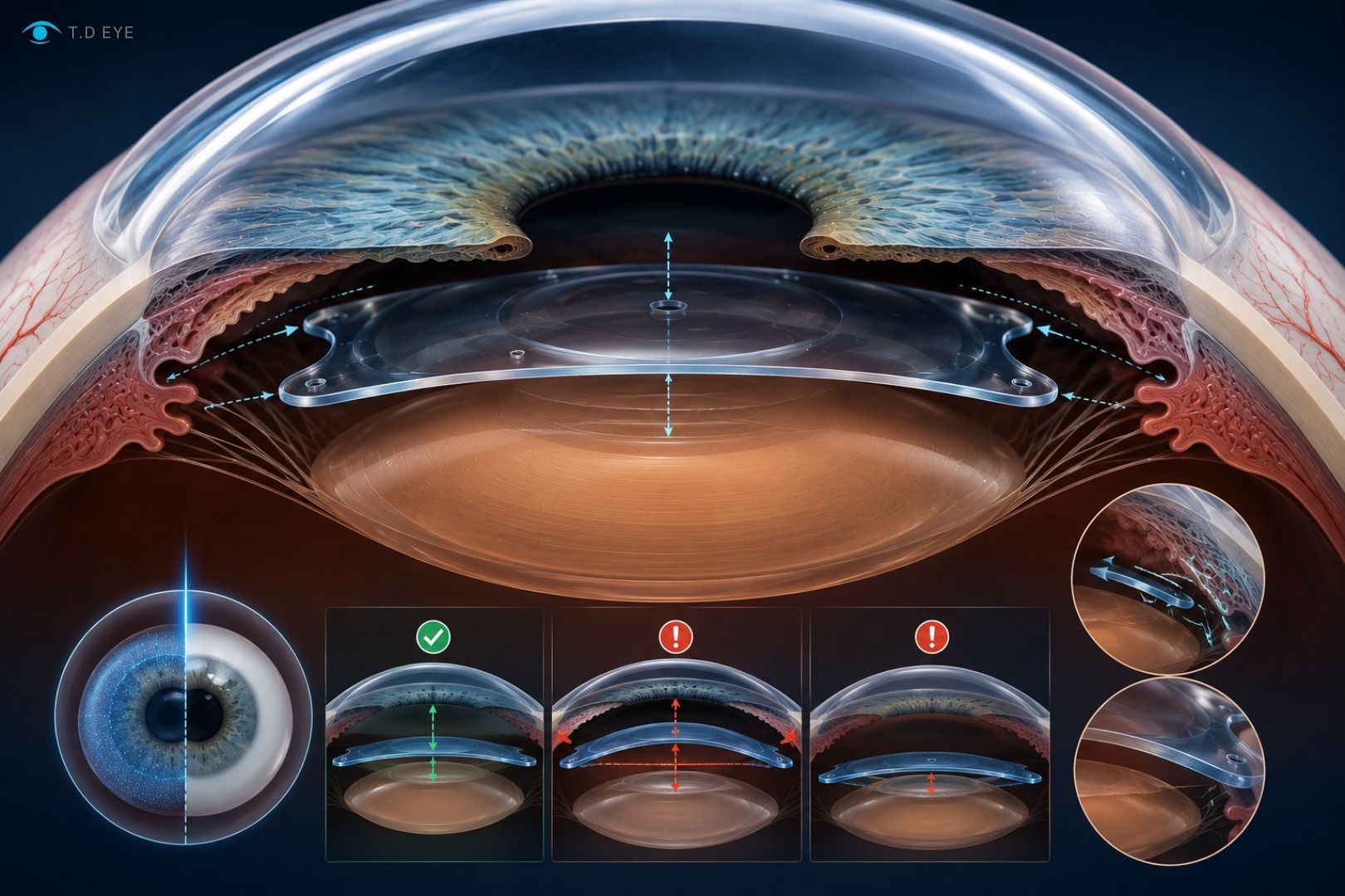
ICL curvature can be understood as the three-dimensional curved shape of the lens once implanted inside the eye, including the design of the anterior surface, posterior surface, and the overall vault of the lens.
This combination determines how far the ICL sits from the crystalline lens, how much vault is created, and how it affects the iris, anterior chamber, anterior chamber angle, as well as aqueous humor circulation.
From an optical perspective, the curvature of the ICL helps redirect light rays so that images focus accurately on the retina. With EVO ICL, the central optical zone includes an additional 0.36 mm central port, also known as KS-AquaPort, which allows aqueous humor to circulate from the posterior chamber to the anterior chamber without requiring peripheral iridotomy as in older ICL generations.
From an anatomical perspective, the posterior curvature of the ICL must sufficiently “fit” within the space between the iris and the natural crystalline lens. If the curvature and size of the ICL are not compatible with the eye’s anatomy, the lens may create excessively low or high vault. This is why Phakic ICL is considered an intraocular device that must be customized for each individual eye.
Why Does ICL Curvature Affect Visual Quality?
Postoperative visual quality after ICL implantation depends on whether the eye’s new optical system is stable, coaxial, and minimally affected by optical aberrations. When the ICL curvature is appropriate, the lens remains balanced within the sulcus, the central optical zone aligns with the pupil axis, and light passing through the eye experiences less scattering.
Conversely, if the ICL is tilted, decentered, or produces abnormal vault, patients may experience glare, halos, reduced contrast sensitivity, or discomfort during night vision.

Why Does ICL Curvature Affect Visual Quality?
A 2025 review on anterior segment OCT reported that tilt below 3–5 degrees and decentration around 0.2–0.3 mm are generally considered stable ranges, whereas decentration exceeding 0.5 mm may be associated with coma, glare, and visual quality disturbances.
| Parameter | Range Commonly Considered Safe | Clinical Significance |
|---|---|---|
| Central ICL Vault | 250–750 µm | Maintains a safe distance between the ICL and crystalline lens |
| Ideal Reference Vault | Approximately 500 µm | Balances the risk of crystalline lens contact and angle narrowing |
| Low Vault | <250 µm | Increases the risk of crystalline lens contact and anterior subcapsular cataract |
| High Vault | >1000 µm | Increases the risk of angle narrowing, glaucoma, and pigment dispersion |
| ICL Tilt | <3–5 degrees | Reduces the risk of optical aberrations and image distortion |
| Decentration | <0.3 mm | Keeps the optical zone stable with the visual axis |
The Relationship Between ICL Curvature and Ocular Structures
The ICL is positioned within a very narrow anatomical space: the iris lies in front, the natural crystalline lens behind, while the outer boundaries relate to the sulcus-to-sulcus distance and the anterior chamber angle. Therefore, ICL curvature cannot be evaluated independently from ocular anatomy.
Compared with corneal refractive procedures, ICL has the advantage of not altering corneal shape. This is especially important in patients with thin corneas, dry eyes, or those unsuitable for corneal laser surgery. EVO ICL documentation emphasizes that LASIK, PRK, and SMILE belong to the group of procedures that reshape the corneal surface, whereas EVO ICL belongs to the category of intraocular lens implantation.
The Relationship Between ICL Curvature and Ocular Structures
Regarding the iris, the curvature and vault of the ICL may slightly push the iris forward. If the vault is excessively high, the iris may be displaced further forward, narrowing the anterior chamber angle and increasing the risk of glaucoma. Regarding the natural crystalline lens, excessively low vault causes the ICL to move closer to or contact the anterior lens capsule, increasing the risk of anterior subcapsular cataract.
| Ocular Structure | How the ICL Interacts | When Parameters Are Inappropriate |
|---|---|---|
| Cornea | Does not alter corneal shape but requires endothelial monitoring | Low endothelial cell density or shallow anterior chamber increases corneal edema risk |
| Iris | The ICL sits behind the iris and may affect iris configuration | High vault may push the iris forward and narrow the angle |
| Crystalline Lens | The ICL sits in front of the crystalline lens and requires a safe distance | Low vault increases cataract risk |
| Anterior Chamber | The anterior chamber space must be sufficiently deep | A shallow anterior chamber reduces the safety margin |
| Anterior Chamber Angle | Acts as the aqueous humor drainage pathway | Angle narrowing may increase intraocular pressure |
| Corneal Endothelium | Must be preserved long-term | Significant endothelial cell loss may affect corneal clarity |
ICL Vault: The Central Parameter of Biological Interaction
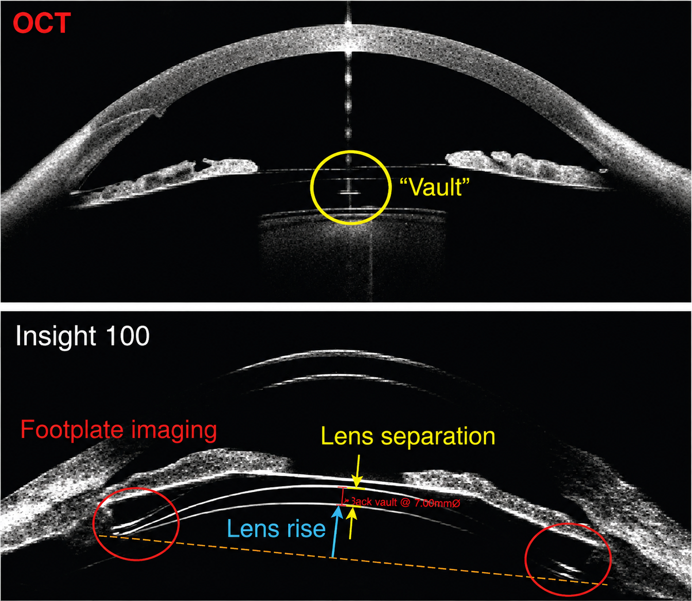
ICL vault is the perpendicular distance between the posterior surface of the ICL and the anterior surface of the natural crystalline lens. This is one of the most important postoperative ICL parameters because it directly reflects whether the lens appropriately “fits” the ocular anatomy.
ICL Vault: The Central Parameter of Biological Interaction
Studies using anterior segment OCT have reported that average vault commonly ranges around 500–600 µm, with a standard deviation of approximately 200 µm. A vault range of 250–750 µm is generally considered the target zone under normal lighting conditions.
Nevertheless, approximately 5–10% of eyes may still fall into the low vault range below 250 µm or the high vault range above 1000 µm even when sizing formulas are used.
| Vault Type | Characteristics | Clinical Significance |
|---|---|---|
| Low Vault | ICL sits too close to the crystalline lens | May increase the risk of anterior subcapsular cataract |
| Ideal Vault | ICL maintains an appropriate distance from the crystalline lens | Balances crystalline lens safety and angle openness |
| High Vault | ICL pushes the iris forward | May cause angle narrowing, glaucoma, glare, or halos |
| Vault Reduction Over Time | Usually associated with age-related crystalline lens changes | Requires periodic postoperative OCT monitoring |
Central Port, Collamer, and Aqueous Humor Circulation
EVO ICL differs from older generations thanks to its 0.36 mm central port. The central port allows aqueous humor to circulate through the center of the lens, thereby reducing the need for preoperative peripheral iridotomy. This design is highly significant because aqueous humor flow is related not only to intraocular pressure but also to metabolic exchange around the crystalline lens.
Collamer material is STAAR’s proprietary collagen-containing polymer described as a biocompatible material. In clinical practice, the softness, flexibility, and compatibility of Collamer help the ICL remain stable inside the eye, but long-term stability still depends heavily on ICL size, curvature, vault, and each patient’s anatomical structure.
Clinical Data from the FDA
In the FDA EVO ICL study involving 629 eyes, uncorrected visual acuity reached 20/40 or better in 99.3% of eyes at 6 months, while 75.9% achieved 20/20 or better. This is important evidence demonstrating the high visual effectiveness of EVO ICL when appropriately indicated and measured.
However, safety data also indicate that ICL surgery requires careful postoperative monitoring. In the EVO ICL study, 125 out of 629 eyes, equivalent to 19.9%, experienced elevated intraocular pressure within the first 1–6 hours after surgery, mainly due to retained viscoelastic material, with pressure returning to normal after 1 day. Three eyes, equivalent to 0.5%, lost more than 30% of endothelial cells after 6 months.
| Indicator in the EVO ICL Study | Result | Clinical Significance |
|---|---|---|
| Number of eyes in the FDA study | 629 eyes | Large enough to evaluate short-term effectiveness |
| Uncorrected visual acuity of 20/40 or better after 6 months | 99.3% | Meets daily visual and driving needs in many cases |
| Uncorrected visual acuity of 20/20 or better after 6 months | 75.9% | Indicates high refractive correction quality |
| Increased intraocular pressure 1–6 hours after surgery | 19.9% | Requires early intraocular pressure monitoring after surgery |
| Endothelial cell loss >30% after 6 months | 0.5% | Emphasizes the role of long-term endothelial monitoring |
| Cataract reported in the study | 0.2% | Low short-term risk but still requires long-term follow-up |
| Additional surgery related to EVO ICL | 1.0% | Position adjustment, replacement, or ICL removal may be needed in a small number of cases |
Why Is ICL Curvature Related to Glare, Halos, and Night Vision?
Glare and halos after ICL implantation are often related to multiple factors at the same time: pupil size in low-light conditions, the optical zone of the ICL, coaxial alignment between the ICL and the visual axis, vault status, residual astigmatism, and each person’s individual neurovisual response.
If the dilated pupil is larger than the effective optical zone, peripheral light may create more scattering, causing patients to see halos around car headlights or bright lights. If the ICL is decentered or tilted, higher-order aberrations may increase, reducing contrast sensitivity, especially at night. FDA documentation also notes that glare and halos may be more noticeable in low-light conditions when the pupil is dilated.
This explains why preoperative ICL eye measurements should not stop at myopia and astigmatism. Doctors need to evaluate pupil size, corneal topography, anterior chamber, anterior chamber angle, ACD, WTW, ATA, STS when appropriate equipment is available, crystalline lens rise, and predicted vault.
Factors That Determine Whether ICL Is Suitable for the Eye
ICL suitability is the result of multiple biological parameters. ACD is one of the fundamental criteria. EVO ICL documentation lists anterior chamber depth below 3.0 mm or an eye shape that is unsuitable for lens implantation as contraindications.
In modern practice, anterior segment OCT helps assess ACD, anterior chamber angle, vault, tilt, decentration, and crystalline lens rise. A 2025 review reported that many surgeons use an ACD threshold from the endothelium to the crystalline lens of over 2.8 mm for myopic ICL, and approximately 3.0 mm for hyperopic ICL or anterior chamber lenses, in order to protect the corneal endothelium.
Comparison Between Phakic ICL and LASIK in Terms of Biological Structure
| Criterion | Phakic ICL | LASIK |
|---|---|---|
| Treatment location | Lens implanted behind the iris and in front of the crystalline lens | Creates a flap and reshapes the cornea |
| Effect on the cornea | Phakic ICL does not cause corneal damage | Alters corneal tissue |
| Suitability for thin corneas | May be an option in many cases | Limited by corneal thickness and shape |
| Dry eye | Less associated with corneal nerve disruption | May cause postoperative dry eye in some patients |
| Factors requiring monitoring | ICL vault, intraocular pressure, endothelium, crystalline lens | Corneal surface, dry eye, refractive stability |
| Future adjustability | The ICL can be removed by a doctor when necessary | Reshaped corneal tissue does not return to its original state |
The Role of OCT and Modern Eye Measurement Technology
Anterior segment OCT has become an important tool in Phakic ICL management because it allows direct visualization of the relationship between the lens and ocular structures. OCT can measure vault, ACD, anterior chamber angle, tilt, and decentration, and it helps detect early risk situations such as an ICL positioned too close to the crystalline lens, excessively high vault, angle narrowing, or abnormal iris configuration.
Intraoperative OCT is also being actively studied. A large study of 574 eyes showed that intraoperative vault measurement strongly correlated with vault at 4 hours and 24 hours after surgery. A 25-eye study showed that iOCT-guided ICL rotation could reduce excessive vault from an average of 1148 µm to 740 µm.
When Is In-Depth Evaluation Needed Before ICL Surgery?
In-depth evaluation is especially necessary when patients have high myopia, high astigmatism, thin corneas, insufficient anterior chamber depth, large pupils in the dark, suspected narrow anterior chamber angles, low corneal endothelial cell density, a history of elevated intraocular pressure, previous eye inflammation, or prior eye surgery.
This group also needs a clear explanation of ICL vault, the risk of early intraocular pressure elevation, the possibility of glare and halos, and the long-term follow-up plan.
In the future, ICL personalization will not rely only on traditional sizing formulas, but will also combine swept-source OCT, 3D anterior chamber analysis, artificial intelligence-based vault prediction, and long-term monitoring of ICL dynamics over time. This direction will help Phakic ICL become increasingly safer, more precise, and better suited to each individual ocular structure.

 03/10/2026











 0916.741.763
 Appointment