 vi
vi 24-Jul-2025
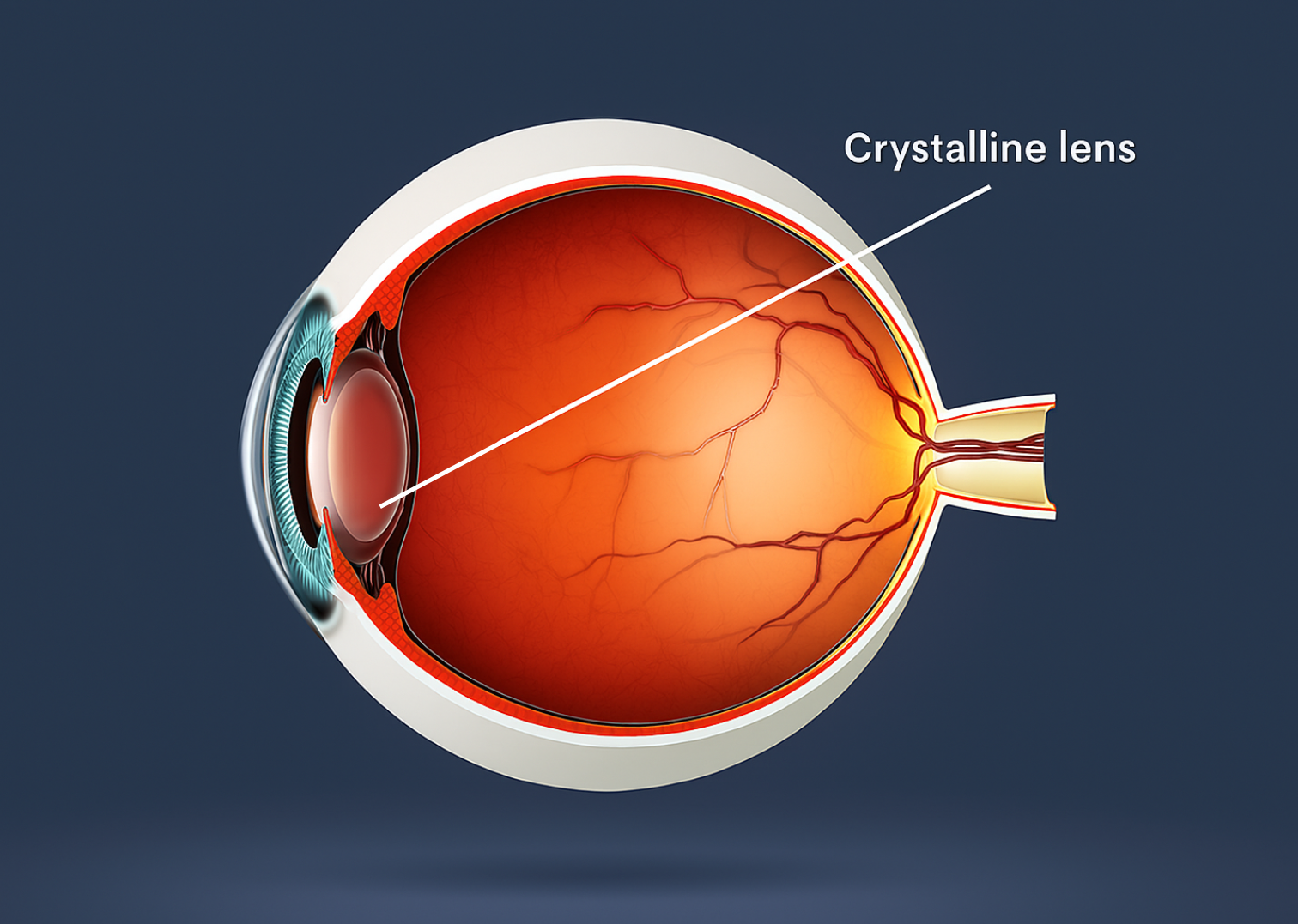
Cornea and Lens in the Optical System of the Eye
The cornea and lens are two vital components of the human eye’s optical system. Together, they manage the direction and focus of light rays, creating a clear and sharp image on the retina. Any disruption in their structure or function can significantly impair vision. This article provides insights into how these two structures operate, their roles in vision, and the modern therapeutic solutions available—especially Phakic ICL, a leading-edge refractive technology adopted by TD Eye.
1/ Cornea – The Eye’s Natural First Lens
The cornea is a dome-shaped, transparent membrane at the front of the eye. As the first surface light hits, it refracts approximately two-thirds of incoming light, making it the eye’s primary focusing structure. Its transparency and curvature are critical to maintaining clear vision.
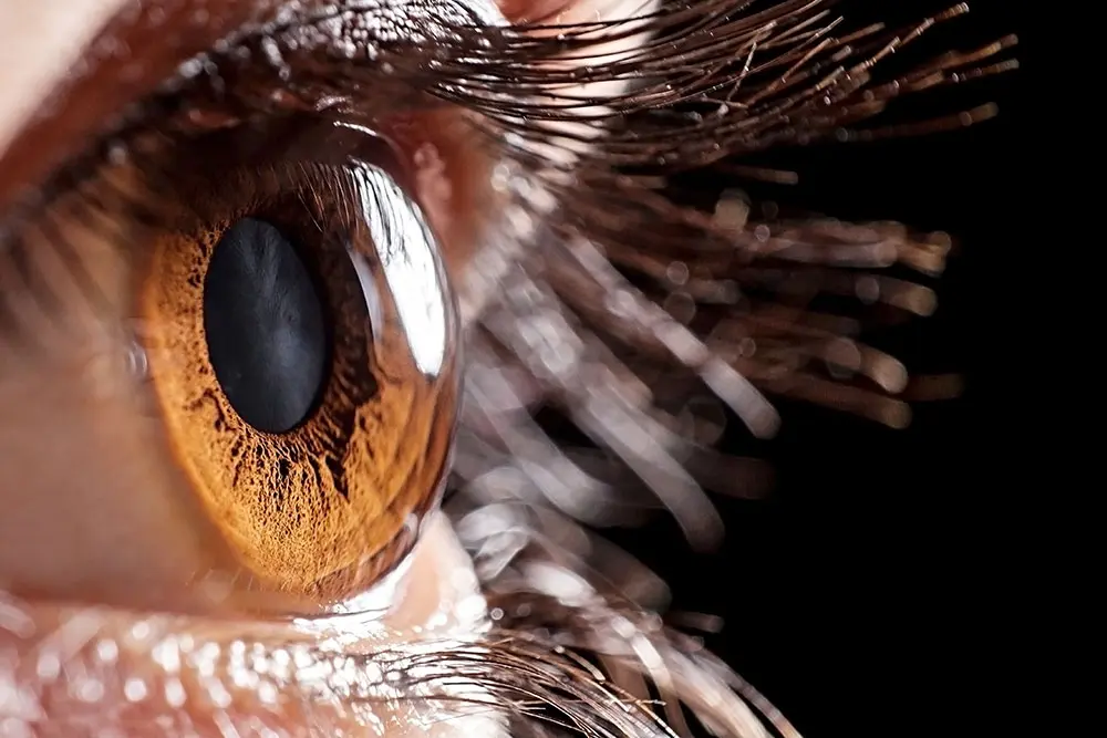
Cornea – the first natural lens that focuses light into the eye.
Notable features include:
- Lack of blood vessels – promotes clear light transmission
- Thousands of nerve endings – highly sensitive to injury and dryness
- Five specialized layers: epithelium, Bowman’s layer, stroma, Descemet’s membrane, and endothelium
Corneal Layers and Their Roles
| Component | Role in Eye Optics |
|---|---|
| Epithelium | Acts as a protective barrier and creates a smooth surface for light refraction |
| Bowman’s Layer | Tough fibrous support – protects against deeper injuries |
| Stroma | Thickest layer – made of organized collagen fibers, defines corneal shape |
| Descemet’s Membrane | Basement layer for endothelial cells, supports the inner structure |
| Endothelium | Maintains dehydration and clarity by actively pumping fluid out |
2/ Lens – The Eye’s Adjustable Zoom
Located just behind the iris, the lens adjusts its curvature dynamically to focus on objects at various distances—near or far. This accommodation is possible due to the action of the ciliary muscles, making them thicker or thinner as needed.
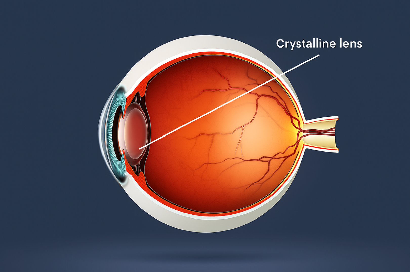
Crystalline lens – structure that adjusts eye focus.
- Continuously reshaped by the ciliary muscles
- Essential for near vision tasks like reading and screen work
- Ages with time – leading to presbyopia (difficulty focusing on near objects) and cataracts
Presbyopia is a natural part of aging that typically begins around age 40, when the eye’s focusing structure loses flexibility and struggles to adjust to nearby objects. Cataracts, another age-related condition, develop when proteins inside this transparent structure clump together, causing cloudiness. Cataract surgery involves replacing the clouded component with an artificial intraocular lens (IOL) to restore clear vision.
3/ How the Eye’s Optical System Works
A healthy optical system channels light through several components:
Cornea (primary refraction) → Aqueous humor (passes light through the pupil) → Lens (adjusts focus) → Retina (converts light to neural signals)
Any failure to precisely converge light onto the retina results in blurred or distorted images. This is the foundation of refractive errors: myopia (nearsightedness), hyperopia (farsightedness), and astigmatism.
Coordination Between the Cornea and Lens
The cornea and lens function as a unit. The cornea provides fixed, strong refractive power, while the lens fine-tunes that refraction depending on object distance. This synergy ensures smooth transitions between distance and near focus.
If either element fails—say, due to irregular corneal shape or a stiffened lens—vision flexibility suffers. Early detection and correction help maintain quality of life.
4/ Corrective Options When Optics Fail
There are three major vision correction strategies:
- Eyeglasses: Widely used, non-invasive, but external and temporary
- Laser Surgery (LASIK, SMILE): Reshapes corneal curvature to improve focus
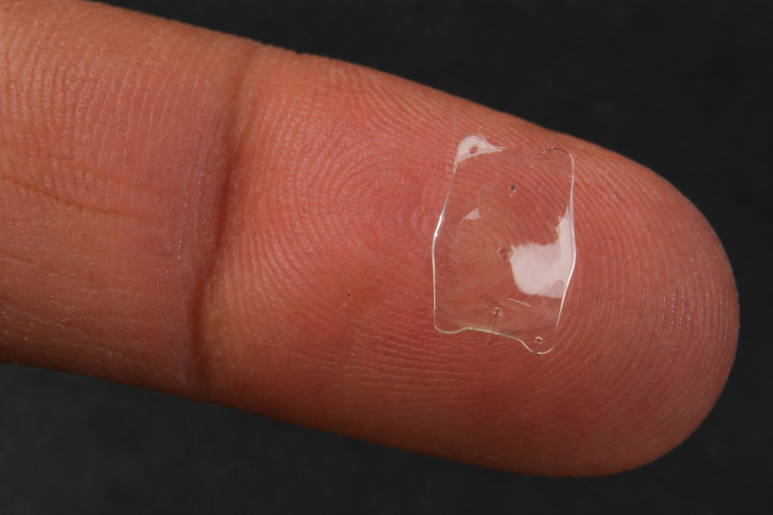
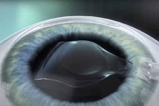
- Phakic ICL: Implantable Collamer Lens placed behind the iris – ideal for high myopia or thin corneas without altering the cornea
5/ Phakic ICL – Advanced Vision Correction That Preserves the Cornea
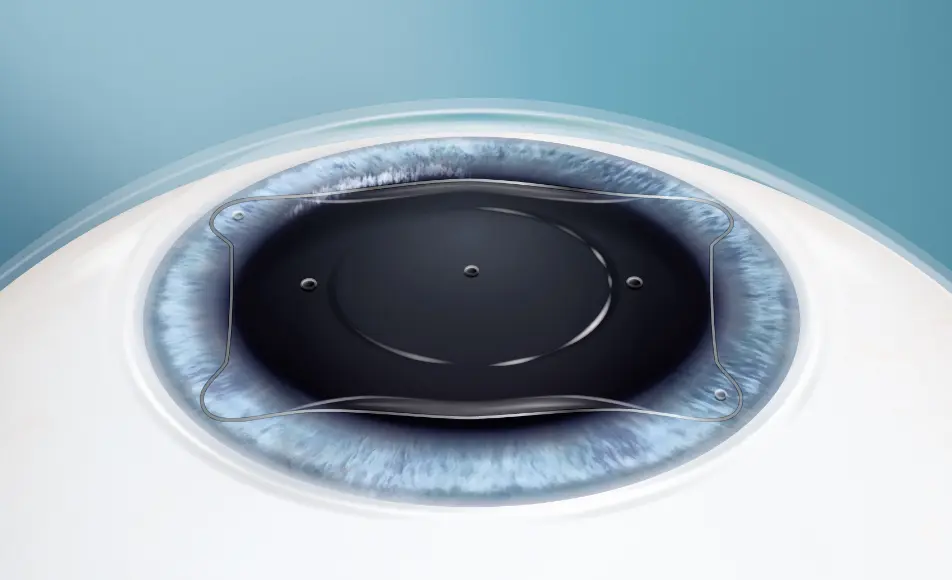
Phakic ICL lens placed behind the iris without corneal damage.
Phakic ICL is a foldable, biocompatible lens implanted between the iris and the natural lens. Unlike laser surgery, it doesn’t cut the cornea, making it reversible and safer for certain patients.
| Criteria | Phakic ICL | Laser Surgery |
|---|---|---|
| Corneal impact | No cutting | Cornea reshaped |
| Reversibility | Yes | No |
| Best for | Thin cornea, severe refractive errors | Moderate prescriptions, thick cornea |
| Dry eye risk | Minimal | Moderate to high |
| Night vision | Stable, minimal glare | Possible halos or starbursts |
Besides long-term clarity, Phakic ICL ensures structural eye integrity and reduces post-operative dryness. It is ideal for those seeking reversible, minimally invasive solutions.
More importantly, Phakic ICL can be considered even in younger patients who are not ready for permanent vision modification but still desire visual freedom. The technology is growing rapidly, supported by clinical studies on safety, patient satisfaction, and long-term outcomes. Combined with topographic corneal mapping and personalized measurements, Phakic ICL procedures at TD Eye are tailored to the individual anatomy and lifestyle of each patient.
6/ Conclusion
The eye’s optical system—led by the cornea and lens—is a masterpiece of biological engineering. Understanding their function, disorders, and correction methods empowers individuals to make informed decisions about eye care.
With innovations like Phakic ICL, patients now have access to tailored solutions that preserve natural eye structure while restoring visual freedom. Early diagnosis, routine exams, and expert consultation are key to maintaining healthy, vibrant eyesight for life.


 0916.741.763
 Appointment