 vi
vi 08-Jul-2025
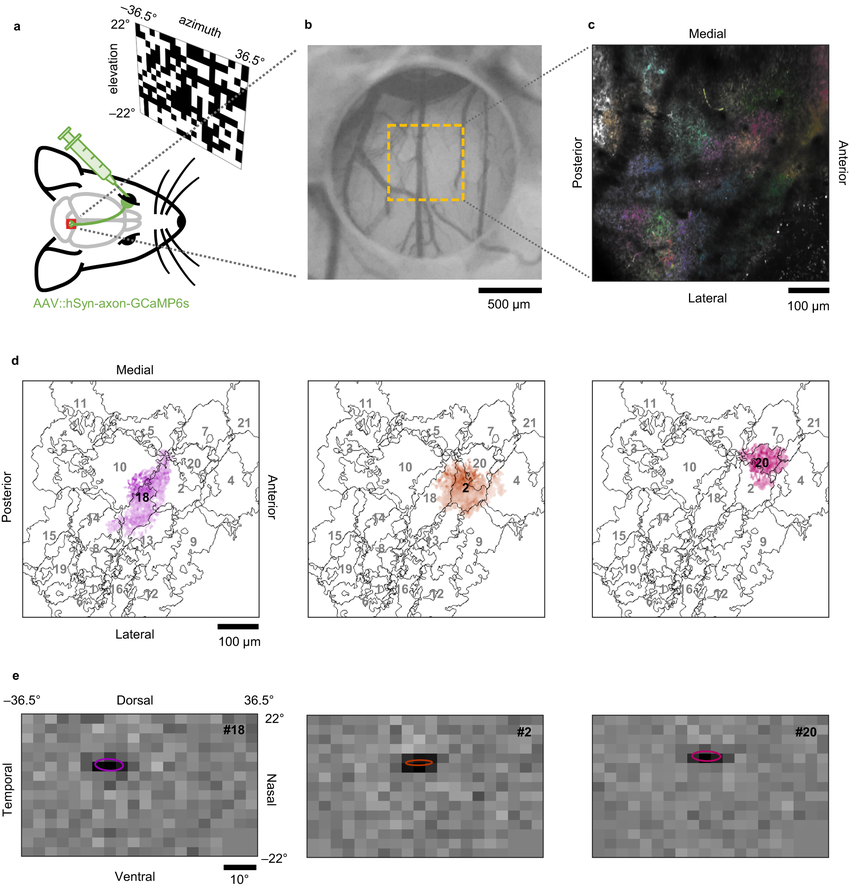
1. At the 2025 International SPECTRALIS Symposium – A Breakthrough in Retinal Neurodiagnostics
At the 2025 International SPECTRALIS Symposium—And Beyond (ISS), held in Heidelberg, Germany on June 13–14, Dr. Balwantray C. Chauhan, PhD, presented his latest research on two-photon microscopy for real-time functional observation of retinal ganglion cells (RGCs) in living subjects.
This technology allows direct, non-invasive monitoring of retinal neuronal activity with high precision, offering promising improvements in assessing the viability of RGCs—critical in conditions such as glaucoma.
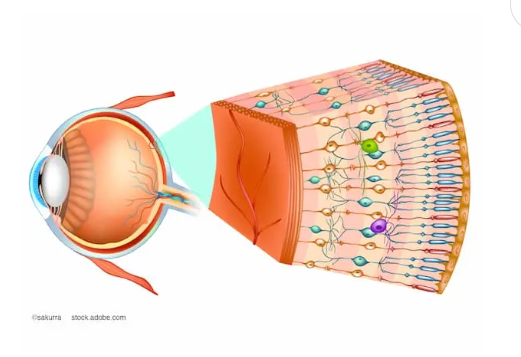
Detailed structure of retinal layers and visual signal transmission.
2. Two-Photon: Addressing the Diagnostic Precision Gap
Dr. Chauhan emphasized that current methods provide only rough estimates of RGC quantity and function. For example, the same RNFL thickness can represent vastly different RGC axon counts across individuals.
This creates a need for single-cell longitudinal tracking at the same site over time. Two-photon technology, in this context, becomes a powerful tool in resolving this gap.
3. How Two-Photon Imaging Works – And Why It’s Safe

Dr. Balwantray C. Chauhan, ophthalmology expert from Dalhousie University, Canada.
A major advantage of this technique is that it causes no observable damage to the retina. According to Dr. Chauhan, his team used low laser power levels (3–8 mW) without detecting any retinal harm in experimental animals.
Although the study was limited to animal models, other research groups have successfully applied two-photon imaging in humans without visible phototoxicity. However, further trials are needed before wider application.
4. How Are Retinal Neurons Tagged with Fluorescent Markers?
A key challenge in RGC in vivo imaging is making the cells “light up” when activated. The team solved this by using adeno-associated virus (AAV) vectors to deliver fluorescent protein genes to retinal ganglion cells.
This enables RGCs to emit unique signals when stimulated by light, allowing precise monitoring of their activity. Notably, AAV vectors do not integrate into the host genome, minimizing genetic safety concerns.
5. Advantages Over Traditional Methods
Unlike ex vivo techniques such as electrophysiology (patch clamp, multi-electrode arrays), which use dissected retinas, two-photon imaging preserves physiological connections and avoids axonal severing.
As a result, it more accurately reflects the natural biological activity of cells in vivo. In contrast, ex vivo signals are limited and may not represent actual cell behavior under normal conditions.
6. Potential Applications in Near-Future Diagnostics
Though not yet widely adopted in clinical settings, this research lays the groundwork for next-generation diagnostic tools in:
- Preclinical glaucoma detection
- Retinal ganglion cell degeneration
- Monitoring effectiveness of neuroprotective treatments
- Quantifying neural recovery over time
7. Conclusion: Measuring Function, Not Just Structure
Dr. Chauhan’s research highlights a core insight in modern ophthalmology: we must assess visual neural function—not just structural imaging.
The technology of two-photon in vivo functional imaging of retinal ganglion cells represents not just a technological breakthrough, but a philosophical shift toward precision medicine and personalized care in vision preservation.


 0916.741.763
 Appointment