 vi
vi 10-Jun-2025
Structure of the Eyeball and Common Refractive Errors
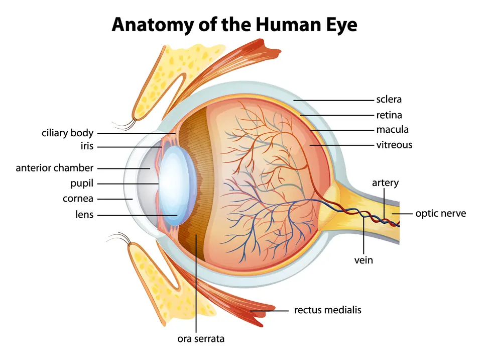
Anatomical Structure of the Human Eye and Its Main Components
In today’s digital age, prolonged screen time can easily strain and damage the eyes. Understanding the structure of the eyeball, how visual focusing works, and the nature of refractive errors is essential for actively protecting and preserving vision.
Anatomy of the Eye and the Mechanism of Clear Vision
The human eye is a sophisticated optical system made up of several tightly coordinated parts. The cornea is the first structure that light encounters, followed by the crystalline lens, which adjusts the focus. Finally, the retina, especially the macula, captures the image and sends visual signals to the brain via the optic nerve.
To see clearly, light must focus precisely on the macula. If it focuses in front of, behind, or off-center from this point, refractive errors occur.
What Are Refractive Errors? Common Types Explained
| Type of Refractive Error | Underlying Mechanism | Main Symptoms |
|---|---|---|
| Nearsightedness (Myopia) | Light focuses in front of the retina | Blurry distance vision |
| Farsightedness (Hyperopia) | Light focuses behind the retina | Blurry near vision |
| Astigmatism | Light focuses unevenly | Distorted or blurred images |
| Presbyopia | Age-related loss of focusing ability | Difficulty reading, need for brighter light |
Causes and Risk Factors
Refractive errors can result from abnormal anatomical structure of the eye—often congenital—or be influenced by genetics and modern lifestyle habits.
| Risk Factor | Associated With |
|---|---|
| Family history (genetics) | Myopia, Astigmatism |
| Poor lighting while working | Progressive myopia |
| Prolonged use of digital devices | Eye strain, poor focusing |
| Aging | Presbyopia |
Common Warning Signs

Simulated Image of Blurred Vision and Glare While Driving at Night
People with refractive errors may experience one or more of the following: blurry vision when reading or looking at distant objects, headaches, eye fatigue after studying or working, or the need to squint to see clearly. Distorted, blurry, or double images—especially at night—are also common signs. Mild symptoms are often overlooked, leading to delayed diagnosis.
Diagnostic Methods
Comprehensive and regular eye exams are essential for early and accurate detection. Common techniques include:
- Visual acuity tests using eye charts and auto-refractors
- Assessment of focusing ability and corneal curvature
- Fundus examination to evaluate the optic nerve and macula
Effective Treatment Options
Depending on the type and severity of the refractive error, treatment may include:
- Eyeglasses or contact lenses: simple and widely used for vision correction
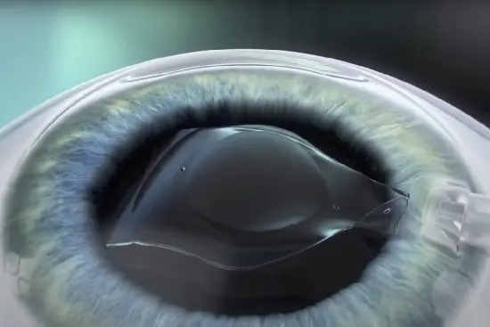
- Refractive surgery (LASIK, SMILE): long-term correction for those with suitable corneal conditions
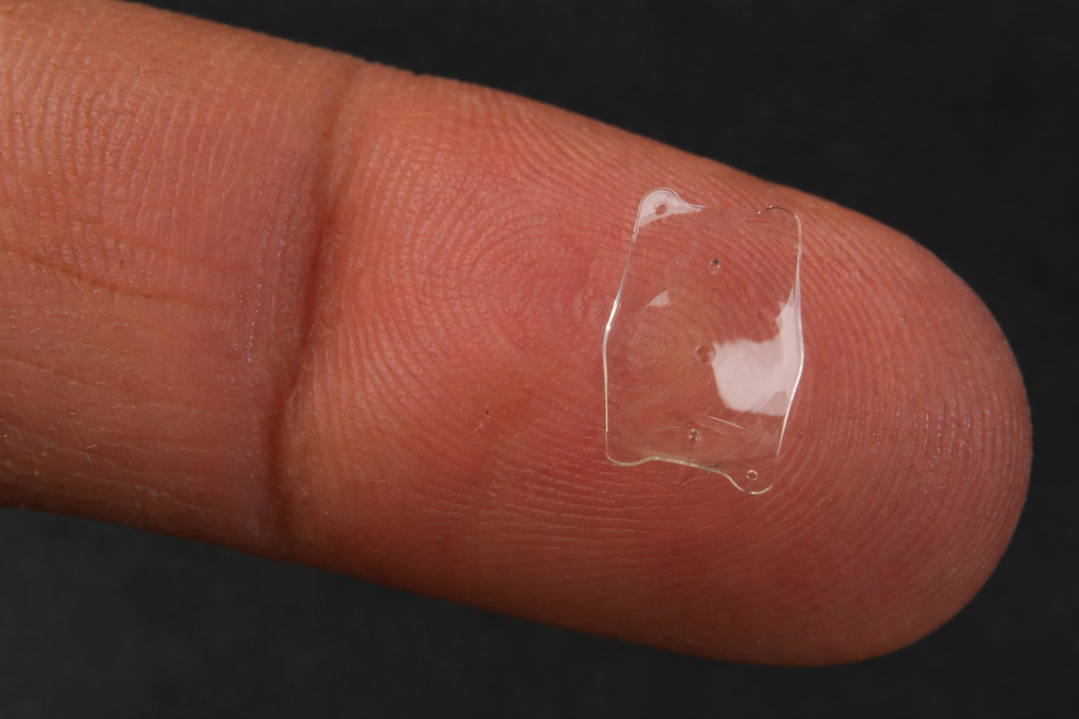
- Phakic ICL lenses: ideal for individuals with thin corneas, dry eyes, or high visual demands
Daily Vision Care Tips
- Follow the 20-20-20 rule: every 20 minutes of screen time, look at something 20 feet away for 20 seconds
- Avoid using mobile devices in low-light conditions
Additionally, support eye health with a diet rich in vitamin A, lutein, and omega-3. Schedule a comprehensive eye exam every 6 months to detect and address issues early.
Refractive errors affect more than just vision—they impact quality of life, learning, and work performance. Proper understanding, timely diagnosis, and appropriate correction are key to maintaining lifelong eye health.


 0916.741.763
 Appointment